A Unique Mdm2-Binding Mode of the 3-Pyrrolin-2-one- and 2-Furanone-Based Antagonists of the p53-Mdm2 Interaction.
Surmiak, E., Twarda-Clapa, A., Zak, K.M., Musielak, B., Tomala, M.D., Kubica, K., Grudnik, P., Madej, M., Jablonski, M., Potempa, J., Kalinowska-Tluscik, J., Domling, A., Dubin, G., Holak, T.A.(2016) ACS Chem Biol 11: 3310-3318
- PubMed: 27709883 Search on PubMed
- DOI: https://doi.org/10.1021/acschembio.6b00596
- Primary Citation Related Structures:
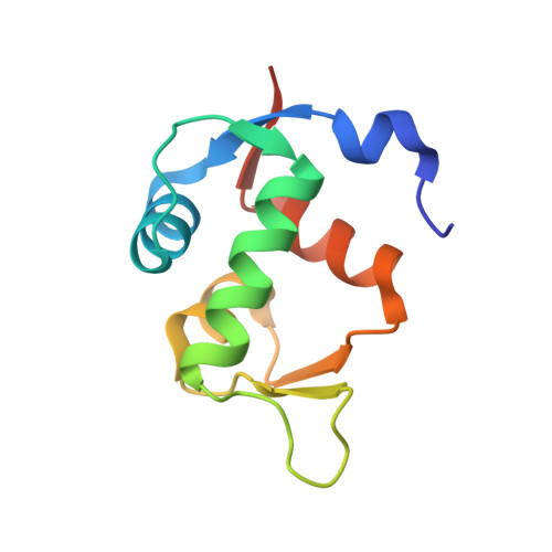
4ZFI, 4ZGK - PubMed Abstract:
The p53 pathway is inactivated in almost all types of cancer by mutations in the p53 encoding gene or overexpression of the p53 negative regulators, Mdm2 and/or Mdmx. Restoration of the p53 function by inhibition of the p53-Mdm2/Mdmx interaction opens up a prospect for a nongenotoxic anticancer therapy. Here, we present the syntheses, activities, and crystal structures of two novel classes of Mdm2-p53 inhibitors that are based on the 3-pyrrolin-2-one and 2-furanone scaffolds. The structures of the complexes formed by these inhibitors and Mdm2 reveal the dimeric protein molecular organization that has not been observed in the small-molecule/Mdm2 complexes described until now. In particular, the 6-chloroindole group does not occupy the usual Trp-23 pocket of Mdm2 but instead is engaged in dimerization. This entirely unique binding mode of the compounds opens new possibilities for optimization of the Mdm2-p53 interaction inhibitors.
- Faculty of Chemistry, Jagiellonian University , Ingardena 3, 30-060 Krakow, Poland.
Organizational Affiliation: