4ZNX

Crystal structure of the Fyn-SH3 domain in complex with the high affinity peptide APP12


Experimental Data Snapshot

  • Method: X-RAY DIFFRACTION
  • Resolution: 2.10 Å
  • R-Value Free: 0.222 
  • R-Value Work: 0.179 
  • R-Value Observed: 0.182 

wwPDB Validation   3D Report Full Report


This is version 1.3 of the entry. See complete history


Literature

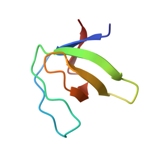
The role of water molecules in the binding of class I and II peptides to the SH3 domain of the Fyn tyrosine kinase.

Camara-Artigas, A.Ortiz-Salmeron, E.Andujar-Sanchez, M.Bacarizo, J.Martin-Garcia, J.M.

(2016) Acta Crystallogr F Struct Biol Commun 72: 707-712

  • DOI: https://doi.org/10.1107/S2053230X16012310
  • Primary Citation of Related Structures:  
    4ZNX

  • PubMed Abstract: 

    Interactions of proline-rich motifs with SH3 domains are present in signal transduction and other important cell processes. Analysis of structural and thermodynamic data suggest a relevant role of water molecules in these protein-protein interactions. To determine whether or not the SH3 domain of the Fyn tyrosine kinase shows the same behaviour, the crystal structures of its complexes with two high-affinity synthetic peptides, VSL12 and APP12, which are class I and II peptides, respectively, have been solved. In the class I complexes two water molecules were found at the binding interface that were not present in the class II complexes. The structures suggest a role of these water molecules in facilitating conformational changes in the SH3 domain to allow the binding of the class I or II peptides. In the third binding pocket these changes modify the cation-π and salt-bridge interactions that determine the affinity of the binding. Comparison of the water molecules involved in the binding of the peptides with previous reported hydration spots suggests a different pattern for the SH3 domains of the Src tyrosine kinase family.


  • Organizational Affiliation

    Department of Chemistry and Physics, University of Almería, Agrifood Campus of International Excellence (ceiA3), Carretera de Sacramento, 04120 Almeria, Spain.


Macromolecules
Find similar proteins by:  (by identity cutoff)  |  3D Structure
Entity ID: 1
MoleculeChains Sequence LengthOrganismDetailsImage
Tyrosine-protein kinase Fyn
A, B, C, D
58Homo sapiensMutation(s): 0 
Gene Names: FYN
EC: 2.7.10.2
UniProt & NIH Common Fund Data Resources
Find proteins for P06241 (Homo sapiens)
Explore P06241 
Go to UniProtKB:  P06241
PHAROS:  P06241
GTEx:  ENSG00000010810 
Entity Groups  
Sequence Clusters30% Identity50% Identity70% Identity90% Identity95% Identity100% Identity
UniProt GroupP06241
Sequence Annotations
Expand
  • Reference Sequence

Find similar proteins by:  Sequence   |   3D Structure  

Entity ID: 2
MoleculeChains Sequence LengthOrganismDetailsImage
APP12
E, F, G, H
12synthetic constructMutation(s): 0 
Entity Groups  
Sequence Clusters30% Identity50% Identity70% Identity90% Identity95% Identity100% Identity
Sequence Annotations
Expand
  • Reference Sequence
Experimental Data & Validation

Experimental Data

  • Method: X-RAY DIFFRACTION
  • Resolution: 2.10 Å
  • R-Value Free: 0.222 
  • R-Value Work: 0.179 
  • R-Value Observed: 0.182 
  • Space Group: P 1 21 1
Unit Cell:
Length ( Å )Angle ( ˚ )
a = 31.658α = 90
b = 76.664β = 94.74
c = 73.175γ = 90
Software Package:
Software NamePurpose
Aimlessdata scaling
PHASERphasing
PHENIXrefinement
PDB_EXTRACTdata extraction
autoPROCdata reduction
autoPROCdata scaling

Structure Validation

View Full Validation Report


Entry History & Funding Information

Deposition Data


Funding OrganizationLocationGrant Number
MINECOSpainBIO2012-39922-C02-02

Revision History  (Full details and data files)

  • Version 1.0: 2016-06-29
    Type: Initial release
  • Version 1.1: 2016-09-14
    Changes: Database references
  • Version 1.2: 2016-09-28
    Changes: Database references
  • Version 1.3: 2024-01-10
    Changes: Data collection, Database references, Refinement description