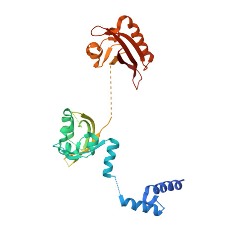
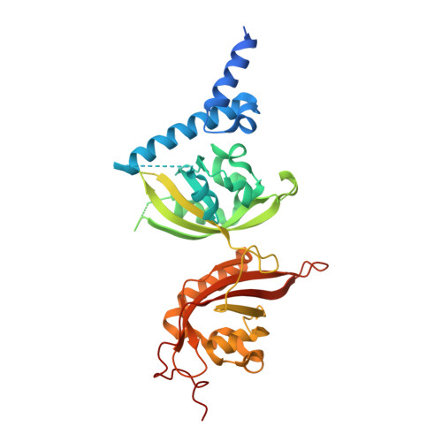
4ZQD

Crystal Structure of the Heterodimeric HIF-2a:ARNT Complex with the Benzoxadiazole Antagonist 0X3


Experimental Data Snapshot

  • Method: X-RAY DIFFRACTION
  • Resolution: 2.87 Å
  • R-Value Free: 0.254 
  • R-Value Work: 0.212 
  • R-Value Observed: 0.218 

Starting Model: experimental
View more details

wwPDB Validation   3D Report Full Report


Ligand Structure Quality Assessment 


This is version 1.4 of the entry. See complete history


Literature

Structural integration in hypoxia-inducible factors.

Wu, D.Potluri, N.Lu, J.Kim, Y.Rastinejad, F.

(2015) Nature 524: 303-308

  • DOI: https://doi.org/10.1038/nature14883
  • Primary Citation of Related Structures:  
    4ZP4, 4ZPH, 4ZPK, 4ZPR, 4ZQD

  • PubMed Abstract: 

    The hypoxia-inducible factors (HIFs) coordinate cellular adaptations to low oxygen stress by regulating transcriptional programs in erythropoiesis, angiogenesis and metabolism. These programs promote the growth and progression of many tumours, making HIFs attractive anticancer targets. Transcriptionally active HIFs consist of HIF-α and ARNT (also called HIF-1β) subunits. Here we describe crystal structures for each of mouse HIF-2α-ARNT and HIF-1α-ARNT heterodimers in states that include bound small molecules and their hypoxia response element. A highly integrated quaternary architecture is shared by HIF-2α-ARNT and HIF-1α-ARNT, wherein ARNT spirals around the outside of each HIF-α subunit. Five distinct pockets are observed that permit small-molecule binding, including PAS domain encapsulated sites and an interfacial cavity formed through subunit heterodimerization. The DNA-reading head rotates, extends and cooperates with a distal PAS domain to bind hypoxia response elements. HIF-α mutations linked to human cancers map to sensitive sites that establish DNA binding and the stability of PAS domains and pockets.


  • Organizational Affiliation

    Metabolic Disease Program, Sanford Burnham Prebys Medical Discovery Institute, Orlando, Florida 32827, USA.


Macromolecules
Find similar proteins by:  (by identity cutoff)  |  3D Structure
Entity ID: 1
MoleculeChains Sequence LengthOrganismDetailsImage
Aryl hydrocarbon receptor nuclear translocator
A, C
384Mus musculusMutation(s): 0 
Gene Names: Arnt
UniProt & NIH Common Fund Data Resources
Find proteins for P53762 (Mus musculus)
Explore P53762 
Go to UniProtKB:  P53762
IMPC:  MGI:88071
Entity Groups  
Sequence Clusters30% Identity50% Identity70% Identity90% Identity95% Identity100% Identity
UniProt GroupP53762
Sequence Annotations
Expand
  • Reference Sequence
Find similar proteins by:  (by identity cutoff)  |  3D Structure
Entity ID: 2
MoleculeChains Sequence LengthOrganismDetailsImage
Endothelial PAS domain-containing protein 1
B, D
360Mus musculusMutation(s): 0 
Gene Names: Epas1Hif2a
UniProt & NIH Common Fund Data Resources
Find proteins for P97481 (Mus musculus)
Explore P97481 
Go to UniProtKB:  P97481
IMPC:  MGI:109169
Entity Groups  
Sequence Clusters30% Identity50% Identity70% Identity90% Identity95% Identity100% Identity
UniProt GroupP97481
Sequence Annotations
Expand
  • Reference Sequence
Small Molecules
Ligands 1 Unique
IDChains Name / Formula / InChI Key2D Diagram3D Interactions
0X3
Query on 0X3

Download Ideal Coordinates CCD File 
E [auth B]N-(3-chloro-5-fluorophenyl)-4-nitro-2,1,3-benzoxadiazol-5-amine
C12 H6 Cl F N4 O3
CDQUJZKBRAFWNG-UHFFFAOYSA-N
Binding Affinity Annotations 
IDSourceBinding Affinity
0X3 BindingDB:  4ZQD Kd: 90 (nM) from 1 assay(s)
IC50: 490 (nM) from 1 assay(s)
Experimental Data & Validation

Experimental Data

  • Method: X-RAY DIFFRACTION
  • Resolution: 2.87 Å
  • R-Value Free: 0.254 
  • R-Value Work: 0.212 
  • R-Value Observed: 0.218 
  • Space Group: P 1
Unit Cell:
Length ( Å )Angle ( ˚ )
a = 49.123α = 89.91
b = 76.334β = 89.99
c = 97.985γ = 73.18
Software Package:
Software NamePurpose
HKL-3000data collection
HKL-3000phasing
PHASERphasing
PHENIXrefinement
PDB_EXTRACTdata extraction
SCALEPACKdata scaling
Cootmodel building
HKL-3000data scaling

Structure Validation

View Full Validation Report


Ligand Structure Quality Assessment 


Entry History 

Deposition Data

Revision History  (Full details and data files)

  • Version 1.0: 2015-08-12
    Type: Initial release
  • Version 1.1: 2015-08-19
    Changes: Database references
  • Version 1.2: 2015-08-26
    Changes: Database references
  • Version 1.3: 2017-11-22
    Changes: Database references, Derived calculations, Refinement description
  • Version 1.4: 2023-09-27
    Changes: Data collection, Database references, Refinement description