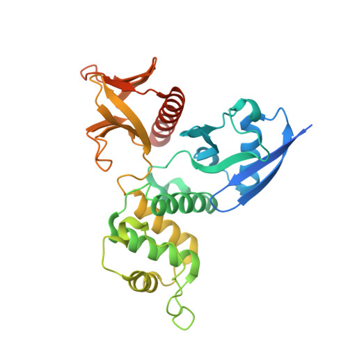
4ZRI | pdb_00004zri

Crystal structure of Merlin-FERM and Lats2


Experimental Data Snapshot

  • Method: X-RAY DIFFRACTION
  • Resolution: 2.70 Å
  • R-Value Free: 
    0.270 (Depositor), 0.282 (DCC) 
  • R-Value Work: 
    0.227 (Depositor), 0.233 (DCC) 
  • R-Value Observed: 
    0.229 (Depositor) 

Starting Model: experimental
View more details

wwPDB Validation 3D Report Full Report

Validation slider image for 4ZRI

This is version 1.4 of the entry. See complete history. 

Literature

Angiomotin binding-induced activation of Merlin/NF2 in the Hippo pathway

Li, Y., Zhou, H., Li, F., Chan, S.W., Lin, Z., Wei, Z., Yang, Z., Guo, F., Lim, C.J., Xing, W., Shen, Y., Hong, W., Long, J., Zhang, M.

(2015) Cell Res 25: 801-817

  • DOI: https://doi.org/10.1038/cr.2015.69
  • Primary Citation Related Structures: 
    4ZRI, 4ZRJ, 4ZRK

  • PubMed Abstract: 

    The tumor suppressor Merlin/NF2 functions upstream of the core Hippo pathway kinases Lats1/2 and Mst1/2, as well as the nuclear E3 ubiquitin ligase CRL4(DCAF1). Numerous mutations of Merlin have been identified in Neurofibromatosis type 2 and other cancer patients. Despite more than two decades of research, the upstream regulator of Merlin in the Hippo pathway remains unknown. Here we show by high-resolution crystal structures that the Lats1/2-binding site on the Merlin FERM domain is physically blocked by Merlin's auto-inhibitory tail. Angiomotin binding releases the auto-inhibition and promotes Merlin's binding to Lats1/2. Phosphorylation of Ser518 outside the Merlin's auto-inhibitory tail does not obviously alter Merlin's conformation, but instead prevents angiomotin from binding and thus inhibits Hippo pathway kinase activation. Cancer-causing mutations clustered in the angiomotin-binding domain impair angiomotin-mediated Merlin activation. Our findings reveal that angiomotin and Merlin respectively interface cortical actin filaments and core kinases in Hippo signaling, and allow construction of a complete Hippo signaling pathway.


  • Organizational Affiliation: 
    • Division of Life Science, State Key Laboratory of Molecular Neuroscience, Hong Kong, China.

Macromolecule Content 

  • Total Structure Weight: 83.09 kDa 
  • Atom Count: 4,975 
  • Modeled Residue Count: 623 
  • Deposited Residue Count: 712 
  • Unique protein chains: 2

Macromolecules

Find similar proteins by:|  3D Structure
Entity ID: 1
MoleculeChains  Sequence LengthOrganismDetailsImage
Merlin
A, B
324Homo sapiensMutation(s): 0 
Gene Names: NF2, SCH
UniProt & NIH Common Fund Data Resources
Find proteins for P35240 (Homo sapiens)
Explore P35240 
Go to UniProtKB:  P35240
PHAROS:  P35240
GTEx:  ENSG00000186575 
Entity Groups
Sequence Clusters30% Identity50% Identity70% Identity90% Identity95% Identity100% Identity
UniProt GroupP35240
Sequence Annotations
Expand
Reference Sequence
Find similar proteins by:|  3D Structure
Entity ID: 2
MoleculeChains  Sequence LengthOrganismDetailsImage
Serine/threonine-protein kinase LATS2
C, D
32Homo sapiensMutation(s): 0 
Gene Names: LATS2, KPM
EC: 2.7.11.1
UniProt & NIH Common Fund Data Resources
Find proteins for Q9NRM7 (Homo sapiens)
Explore Q9NRM7 
Go to UniProtKB:  Q9NRM7
PHAROS:  Q9NRM7
GTEx:  ENSG00000150457 
Entity Groups
Sequence Clusters30% Identity50% Identity70% Identity90% Identity95% Identity100% Identity
UniProt GroupQ9NRM7
Sequence Annotations
Expand
Reference Sequence

Experimental Data & Validation

Experimental Data

  • Method: X-RAY DIFFRACTION
  • Resolution: 2.70 Å
  • R-Value Free:  0.270 (Depositor), 0.282 (DCC) 
  • R-Value Work:  0.227 (Depositor), 0.233 (DCC) 
  • R-Value Observed: 0.229 (Depositor) 
Space Group: P 1 21 1
Unit Cell:
Length ( Å )Angle ( ˚ )
a = 62.962α = 90
b = 100.549β = 105.85
c = 65.03γ = 90
Software Package:
Software NamePurpose
PHENIXrefinement
Blu-Icedata collection
HKL-2000data scaling
PHASERphasing

Structure Validation

View Full Validation Report


Entry History 

Deposition Data

Revision History  (Full details and data files)

  • Version 1.0: 2015-06-17
    Type: Initial release
  • Version 1.1: 2015-07-15
    Changes: Database references
  • Version 1.2: 2015-09-16
    Changes: Other
  • Version 1.3: 2023-11-08
    Changes: Data collection, Database references, Derived calculations, Refinement description
  • Version 1.4: 2024-11-13
    Changes: Structure summary