4ZS7

Structural mimicry of receptor interaction by antagonistic IL-6 antibodies


Experimental Data Snapshot

  • Method: X-RAY DIFFRACTION
  • Resolution: 2.93 Å
  • R-Value Free: 0.293 
  • R-Value Work: 0.261 
  • R-Value Observed: 0.265 

wwPDB Validation   3D Report Full Report


This is version 1.2 of the entry. See complete history


Literature

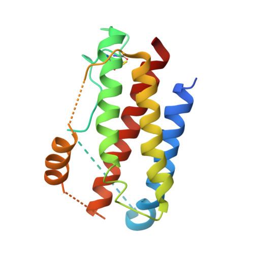
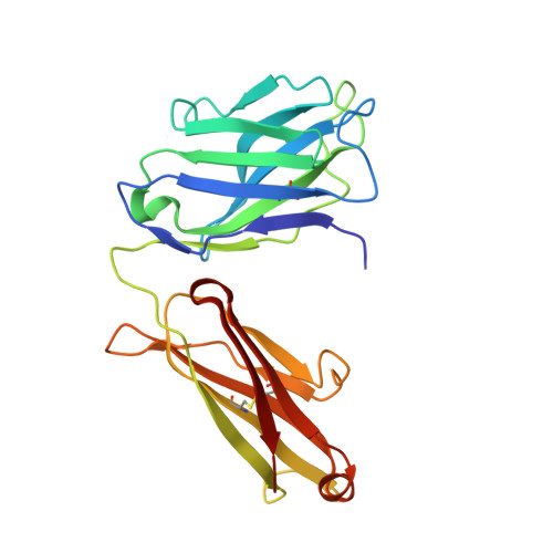
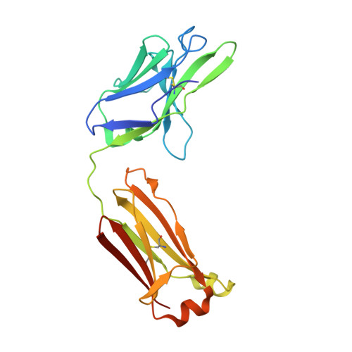
Structural Mimicry of Receptor Interaction by Antagonistic Interleukin-6 (IL-6) Antibodies.

Blanchetot, C.De Jonge, N.Desmyter, A.Ongenae, N.Hofman, E.Klarenbeek, A.Sadi, A.Hultberg, A.Kretz-Rommel, A.Spinelli, S.Loris, R.Cambillau, C.de Haard, H.

(2016) J Biol Chem 291: 13846-13854

  • DOI: https://doi.org/10.1074/jbc.M115.695528
  • Primary Citation of Related Structures:  
    4ZS7

  • PubMed Abstract: 

    Interleukin 6 plays a key role in mediating inflammatory reactions in autoimmune diseases and cancer, where it is also involved in metastasis and tissue invasion. Neutralizing antibodies against IL-6 and its receptor have been approved for therapeutic intervention or are in advanced stages of clinical development. Here we describe the crystal structures of the complexes of IL-6 with two Fabs derived from conventional camelid antibodies that antagonize the interaction between the cytokine and its receptor. The x-ray structures of these complexes provide insights into the mechanism of neutralization by the two antibodies and explain the very high potency of one of the antibodies. It effectively competes for binding to the cytokine with IL-6 receptor (IL-6R) by using side chains of two CDR residues filling the site I cavities of IL-6, thus mimicking the interactions of Phe(229) and Phe(279) of IL-6R. In the first antibody, a HCDR3 tryptophan binds similarly to hot spot residue Phe(279) Mutation of this HCDR3 Trp residue into any other residue except Tyr or Phe significantly weakens binding of the antibody to IL-6, as was also observed for IL-6R mutants of Phe(279) In the second antibody, the side chain of HCDR3 valine ties into site I like IL-6R Phe(279), whereas a LCDR1 tyrosine side chain occupies a second cavity within site I and mimics the interactions of IL-6R Phe(229).


  • Organizational Affiliation

    From argenx, 9052 Zwijnaarde, Belgium.


Macromolecules
Find similar proteins by:  (by identity cutoff)  |  3D Structure
Entity ID: 1
MoleculeChains Sequence LengthOrganismDetailsImage
Interleukin-6171Homo sapiensMutation(s): 0 
Gene Names: IL6IFNB2
UniProt & NIH Common Fund Data Resources
Find proteins for P05231 (Homo sapiens)
Explore P05231 
Go to UniProtKB:  P05231
PHAROS:  P05231
GTEx:  ENSG00000136244 
Entity Groups  
Sequence Clusters30% Identity50% Identity70% Identity90% Identity95% Identity100% Identity
UniProt GroupP05231
Sequence Annotations
Expand
  • Reference Sequence
Find similar proteins by:  (by identity cutoff)  |  3D Structure
Entity ID: 2
MoleculeChains Sequence LengthOrganismDetailsImage
Llama Fab fragment 68F2 heavy chainB [auth H]222Lama glamaMutation(s): 0 
Entity Groups  
Sequence Clusters30% Identity50% Identity70% Identity90% Identity95% Identity100% Identity
Sequence Annotations
Expand
  • Reference Sequence
Find similar proteins by:  (by identity cutoff)  |  3D Structure
Entity ID: 3
MoleculeChains Sequence LengthOrganismDetailsImage
Llama Fab fragment 68F2 light chainC [auth L]216Lama glamaMutation(s): 0 
Entity Groups  
Sequence Clusters30% Identity50% Identity70% Identity90% Identity95% Identity100% Identity
Sequence Annotations
Expand
  • Reference Sequence
Experimental Data & Validation

Experimental Data

  • Method: X-RAY DIFFRACTION
  • Resolution: 2.93 Å
  • R-Value Free: 0.293 
  • R-Value Work: 0.261 
  • R-Value Observed: 0.265 
  • Space Group: P 1 2 1
Unit Cell:
Length ( Å )Angle ( ˚ )
a = 46.55α = 90
b = 92.86β = 103.93
c = 84.63γ = 90
Software Package:
Software NamePurpose
BUSTERrefinement
XDSdata reduction
XSCALEdata scaling
MOLREPphasing

Structure Validation

View Full Validation Report


Entry History 

Revision History  (Full details and data files)

  • Version 1.0: 2016-05-04
    Type: Initial release
  • Version 1.1: 2016-05-11
    Changes: Database references
  • Version 1.2: 2016-07-06
    Changes: Database references