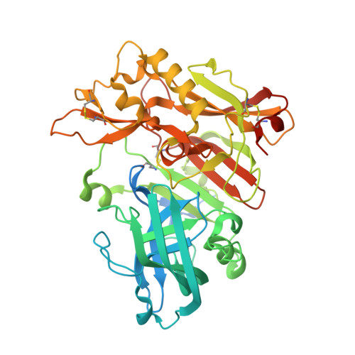
4ZSQ

BACE crystal structure with tricyclic aminothiazine inhibitor


Experimental Data Snapshot

  • Method: X-RAY DIFFRACTION
  • Resolution: 2.30 Å
  • R-Value Free: 0.258 
  • R-Value Work: 0.202 
  • R-Value Observed: 0.203 

wwPDB Validation   3D Report Full Report


Ligand Structure Quality Assessment 

Created with Raphaël 2.3.0Worse 01 BetterLigand structure goodness of fit to experimental dataBest fitted 4RXClick on this verticalbar to view details

This is version 1.2 of the entry. See complete history


Literature

Preparation and biological evaluation of conformationally constrained BACE1 inhibitors.

Winneroski, L.L.Schiffler, M.A.Erickson, J.A.May, P.C.Monk, S.A.Timm, D.E.Audia, J.E.Beck, J.P.Boggs, L.N.Borders, A.R.Boyer, R.D.Brier, R.A.Hudziak, K.J.Klimkowski, V.J.Garcia Losada, P.Mathes, B.M.Stout, S.L.Watson, B.M.Mergott, D.J.

(2015) Bioorg Med Chem 23: 3260-3268

  • DOI: https://doi.org/10.1016/j.bmc.2015.04.062
  • Primary Citation of Related Structures:  
    4ZSM, 4ZSP, 4ZSQ, 4ZSR

  • PubMed Abstract: 

    The BACE1 enzyme is a key target for Alzheimer's disease. During our BACE1 research efforts, fragment screening revealed that bicyclic thiazine 3 had low millimolar activity against BACE1. Analysis of the co-crystal structure of 3 suggested that potency could be increased through extension toward the S3 pocket and through conformational constraint of the thiazine core. Pursuit of S3-binding groups produced low micromolar inhibitor 6, which informed the S3-design for constrained analogs 7 and 8, themselves prepared via independent, multi-step synthetic routes. Biological characterization of BACE inhibitors 6-8 is described.


  • Organizational Affiliation

    Lilly Research Laboratories, A Division of Eli Lilly & Co., Lilly Corporate Center, Indianapolis, IN 46285, USA.


Macromolecules
Find similar proteins by:  (by identity cutoff)  |  3D Structure
Entity ID: 1
MoleculeChains Sequence LengthOrganismDetailsImage
Beta-secretase 1
A, B
442Homo sapiensMutation(s): 0 
Gene Names: BACE1BACEKIAA1149
EC: 3.4.23.46
UniProt & NIH Common Fund Data Resources
Find proteins for P56817 (Homo sapiens)
Explore P56817 
Go to UniProtKB:  P56817
PHAROS:  P56817
GTEx:  ENSG00000186318 
Entity Groups  
Sequence Clusters30% Identity50% Identity70% Identity90% Identity95% Identity100% Identity
UniProt GroupP56817
Sequence Annotations
Expand
  • Reference Sequence
Experimental Data & Validation

Experimental Data

  • Method: X-RAY DIFFRACTION
  • Resolution: 2.30 Å
  • R-Value Free: 0.258 
  • R-Value Work: 0.202 
  • R-Value Observed: 0.203 
  • Space Group: P 21 21 21
Unit Cell:
Length ( Å )Angle ( ˚ )
a = 86.031α = 90
b = 92.629β = 90
c = 132.031γ = 90
Software Package:
Software NamePurpose
REFMACrefinement
MOSFLMdata reduction
SCALAdata scaling
REFMACphasing

Structure Validation

View Full Validation Report


Ligand Structure Quality Assessment 

Created with Raphaël 2.3.0Worse 01 BetterLigand structure goodness of fit to experimental dataBest fitted 4RXClick on this verticalbar to view details

Entry History 

Deposition Data

  • Released Date: 2015-06-10 
  • Deposition Author(s): Timm, D.E.

Revision History  (Full details and data files)

  • Version 1.0: 2015-06-10
    Type: Initial release
  • Version 1.1: 2015-06-17
    Changes: Database references
  • Version 1.2: 2024-11-20
    Changes: Data collection, Database references, Derived calculations, Source and taxonomy, Structure summary