4ZSW | pdb_00004zsw

Pig Brain GABA-AT inactivated by (E)-(1S,3S)-3-Amino-4-fluoromethylenyl-1-cyclopentanoic acid


Experimental Data Snapshot

  • Method: X-RAY DIFFRACTION
  • Resolution: 1.70 Å
  • R-Value Free: 
    0.195 (Depositor), 0.197 (DCC) 
  • R-Value Work: 
    0.158 (Depositor), 0.162 (DCC) 
  • R-Value Observed: 
    0.159 (Depositor) 

wwPDB Validation 3D Report Full Report

Validation slider image for 4ZSW

Ligand Structure Quality Assessment 


This is version 2.1 of the entry. See complete history

Literature

Mechanism of Inactivation of GABA Aminotransferase by (E)- and (Z)-(1S,3S)-3-Amino-4-fluoromethylenyl-1-cyclopentanoic Acid.

Lee, H.Le, H.V.Wu, R.Doud, E.Sanishvili, R.Kellie, J.F.Compton, P.D.Pachaiyappan, B.Liu, D.Kelleher, N.L.Silverman, R.B.

(2015) ACS Chem Biol 10: 2087-2098

  • DOI: https://doi.org/10.1021/acschembio.5b00212
  • Primary Citation Related Structures: 
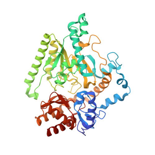
    4ZSW, 4ZSY

  • PubMed Abstract: 

    When γ-aminobutyric acid (GABA), the major inhibitory neurotransmitter in the mammalian central nervous system, falls below a threshold level, seizures occur. One approach to raise GABA concentrations is to inhibit GABA aminotransferase (GABA-AT), a pyridoxal 5'-phosphate-dependent enzyme that degrades GABA. We have previously developed (1S,3S)-3-amino-4-difluoromethylene-1-cyclopentanoic acid (CPP-115), which is 186 times more efficient in inactivating GABA-AT than vigabatrin, the only FDA-approved inactivator of GABA-AT. We also developed (E)- and (Z)-(1S,3S)-3-amino-4-fluoromethylenyl-1-cyclopentanoic acid (1 and 2, respectively), monofluorinated analogs of CPP-115, which are comparable to vigabatrin in inactivating GABA-AT. Here, we report the mechanism of inactivation of GABA-AT by 1 and 2. Both produce a metabolite that induces disruption of the Glu270-Arg445 salt bridge to accommodate interaction between the metabolite formyl group and Arg445. This is the second time that Arg445 has interacted with a ligand and is involved in GABA-AT inactivation, thereby confirming the importance of Arg445 in future inactivator design.


  • Organizational Affiliation
    • Departments of Chemistry and Molecular Biosciences, Chemistry of Life Processes Institute, and the Center for Molecular Innovation and Drug Discovery, Northwestern University , Evanston, Illinois 60208, United States.

Macromolecule Content 

  • Total Structure Weight: 211.17 kDa 
  • Atom Count: 16,678 
  • Modeled Residue Count: 1,844 
  • Deposited Residue Count: 1,844 
  • Unique protein chains: 1

Macromolecules

Find similar proteins by:|  3D Structure
Entity ID: 1
MoleculeChains  Sequence LengthOrganismDetailsImage
4-aminobutyrate aminotransferase, mitochondrial
A, B, C, D
461Sus scrofaMutation(s): 0 
Gene Names: ABATGABAT
EC: 2.6.1.19 (PDB Primary Data), 2.6.1.22 (PDB Primary Data)
UniProt
Find proteins for P80147 (Sus scrofa)
Explore P80147 
Go to UniProtKB:  P80147
Entity Groups
Sequence Clusters30% Identity50% Identity70% Identity90% Identity95% Identity100% Identity
UniProt GroupP80147
Sequence Annotations
Expand
Reference Sequence

Small Molecules

Ligands 4 Unique
IDChains Name / Formula / InChI Key2D Diagram3D Interactions
RW2

Query on RW2


Download:Ideal Coordinates CCD File
F [auth A],
H [auth B],
L [auth C],
M [auth D]
(1S)-4-[({3-hydroxy-2-methyl-5-[(phosphonooxy)methyl]pyridin-4-yl}methyl)amino]cyclopent-3-ene-1,3-dicarboxylic acid
C15 H19 N2 O9 P
VNQKGJPXVHNUEY-QMMMGPOBSA-N
FES

Query on FES


Download:Ideal Coordinates CCD File
E [auth A],
I [auth C]
FE2/S2 (INORGANIC) CLUSTER
Fe2 S2
NIXDOXVAJZFRNF-UHFFFAOYSA-N
GOL

Query on GOL


Download:Ideal Coordinates CCD File
G [auth B],
J [auth C]
GLYCEROL
C3 H8 O3
PEDCQBHIVMGVHV-UHFFFAOYSA-N
ACT

Query on ACT


Download:Ideal Coordinates CCD File
K [auth C]ACETATE ION
C2 H3 O2
QTBSBXVTEAMEQO-UHFFFAOYSA-M

Experimental Data & Validation

Experimental Data

  • Method: X-RAY DIFFRACTION
  • Resolution: 1.70 Å
  • R-Value Free:  0.195 (Depositor), 0.197 (DCC) 
  • R-Value Work:  0.158 (Depositor), 0.162 (DCC) 
  • R-Value Observed: 0.159 (Depositor) 
Space Group: P 1 21 1
Unit Cell:
Length ( Å )Angle ( ˚ )
a = 69.601α = 90
b = 227.614β = 108.97
c = 71.343γ = 90
Software Package:
Software NamePurpose
SCALEPACKdata scaling
PHENIXrefinement
PDB_EXTRACTdata extraction
HKL-2000data reduction
PHASERphasing

Structure Validation

View Full Validation Report


Ligand Structure Quality Assessment 


Entry History 

& Funding Information

Deposition Data


Funding OrganizationLocationGrant Number
National Institutes of Health/National Institute of General Medical Sciences (NIH/NIGMS)United StatesGM066132
National Institutes of Health/National Institute on Drug Abuse (NIH/NIDA)United StatesDA030604
National Institutes of Health/National Institute of General Medical Sciences (NIH/NIGMS)United StatesGM067725
National Institutes of Health/National Cancer Institute (NIH/NCI)United StatesACB-12002
National Institutes of Health/National Institute of General Medical Sciences (NIH/NIGMS)United StatesAGM-12006
Department of Energy (DOE, United States)United StatesDE-AC02-06CH11357

Revision History  (Full details and data files)

  • Version 1.0: 2015-07-08
    Type: Initial release
  • Version 1.1: 2015-09-30
    Changes: Database references
  • Version 1.2: 2017-09-20
    Changes: Advisory, Author supporting evidence, Derived calculations
  • Version 1.3: 2019-12-04
    Changes: Advisory, Author supporting evidence
  • Version 1.4: 2019-12-11
    Changes: Author supporting evidence
  • Version 2.0: 2021-01-13
    Changes: Advisory, Atomic model, Data collection, Derived calculations, Non-polymer description, Structure summary
  • Version 2.1: 2024-03-06
    Changes: Advisory, Data collection, Database references