4AC9 | pdb_00004ac9

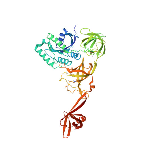
CRYSTAL STRUCTURE OF TRANSLATION ELONGATION FACTOR SELB FROM METHANOCOCCUS MARIPALUDIS IN COMPLEX WITH GDP


Experimental Data Snapshot

  • Method: X-RAY DIFFRACTION
  • Resolution: 3.03 Å
  • R-Value Free: 
    0.222 (Depositor), 0.232 (DCC) 
  • R-Value Work: 
    0.190 (Depositor), 0.200 (DCC) 
  • R-Value Observed: 
    0.191 (Depositor) 

wwPDB Validation 3D Report Full Report

Validation slider image for 4AC9

Ligand Structure Quality Assessment 


This is version 2.0 of the entry. See complete history

Literature

Selenocysteine tRNA-Specific Elongation Factor Selb is a Structural Chimaera of Elongation and Initiation Factors.

Leibundgut, M.Frick, C.Thanbichler, M.Bock, A.Ban, N.

(2005) EMBO J 24: 11

  • DOI: https://doi.org/10.1038/sj.emboj.7600505
  • Primary Citation Related Structures: 
    4AC9, 4ACA, 4ACB

  • PubMed Abstract: 

    In all three kingdoms of life, SelB is a specialized translation elongation factor responsible for the cotranslational incorporation of selenocysteine into proteins by recoding of a UGA stop codon in the presence of a downstream mRNA hairpin loop. Here, we present the X-ray structures of SelB from the archaeon Methanococcus maripaludis in the apo-, GDP- and GppNHp-bound form and use mutational analysis to investigate the role of individual amino acids in its aminoacyl-binding pocket. All three SelB structures reveal an EF-Tu:GTP-like domain arrangement. Upon binding of the GTP analogue GppNHp, a conformational change of the Switch 2 region in the GTPase domain leads to the exposure of SelB residues involved in clamping the 5' phosphate of the tRNA. A conserved extended loop in domain III of SelB may be responsible for specific interactions with tRNA(Sec) and act as a ruler for measuring the extra long acceptor arm. Domain IV of SelB adopts a beta barrel fold and is flexibly tethered to domain III. The overall domain arrangement of SelB resembles a 'chalice' observed so far only for initiation factor IF2/eIF5B. In our model of SelB bound to the ribosome, domain IV points towards the 3' mRNA entrance cleft ready to interact with the downstream secondary structure element.


  • Organizational Affiliation
    • Institut für Molekularbiologie und Biophysik, Eidgenössische Technische Hochschule Zürich, Zürich, Switzerland.

Macromolecule Content 

  • Total Structure Weight: 221.79 kDa 
  • Atom Count: 14,628 
  • Modeled Residue Count: 1,842 
  • Deposited Residue Count: 1,928 
  • Unique protein chains: 1

Macromolecules

Find similar proteins by:|  3D Structure
Entity ID: 1
MoleculeChains  Sequence LengthOrganismDetailsImage
MJ0495-LIKE PROTEIN
A, B, C, D
482Methanococcus maripaludisMutation(s): 0 
UniProt
Find proteins for Q8J307 (Methanococcus maripaludis)
Explore Q8J307 
Go to UniProtKB:  Q8J307
Entity Groups
Sequence Clusters30% Identity50% Identity70% Identity90% Identity95% Identity100% Identity
UniProt GroupQ8J307
Sequence Annotations
Expand
Reference Sequence

Small Molecules

Ligands 6 Unique
IDChains Name / Formula / InChI Key2D Diagram3D Interactions
GDP

Query on GDP


Download:Ideal Coordinates CCD File
E [auth A],
K [auth B],
U [auth C]
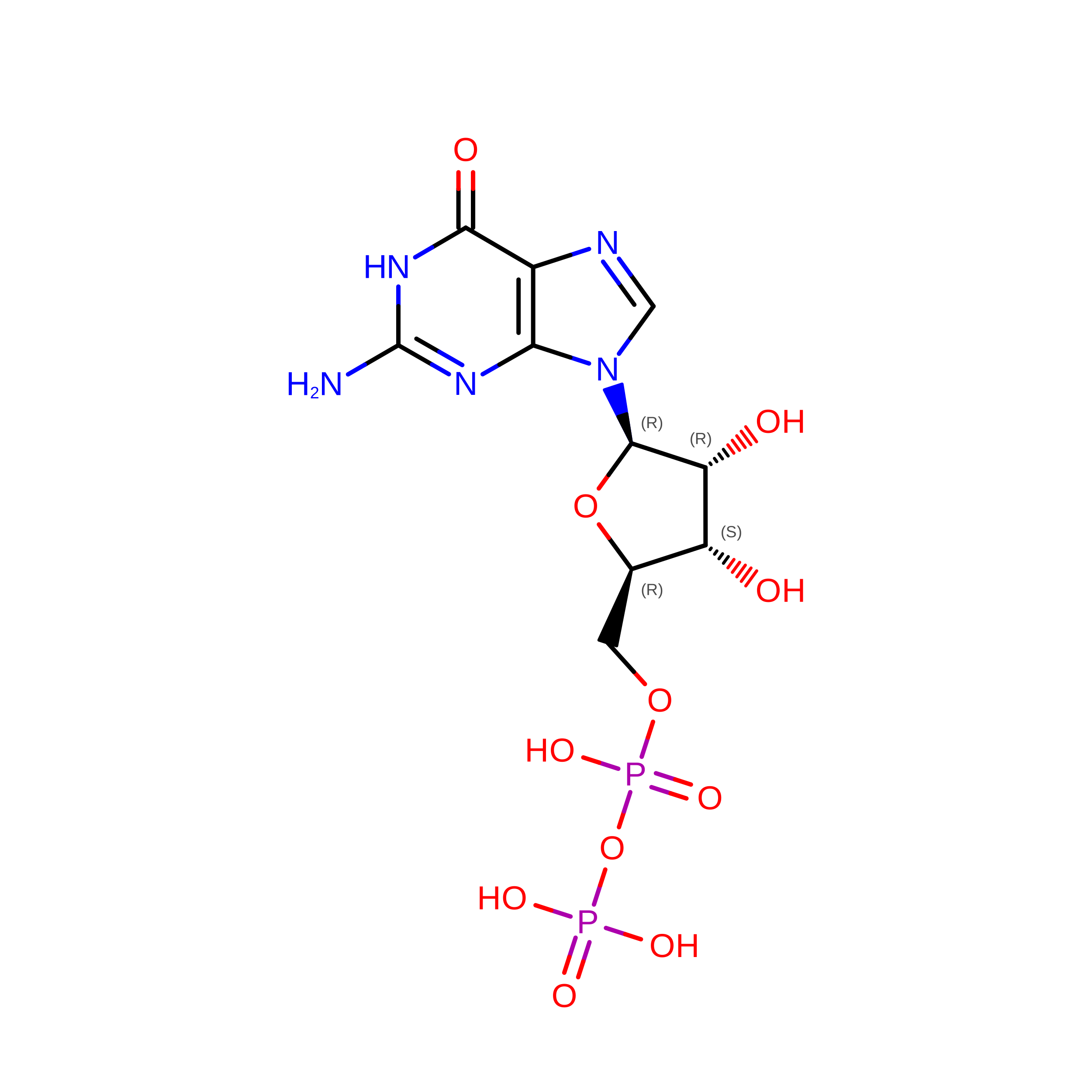
GUANOSINE-5'-DIPHOSPHATE
C10 H15 N5 O11 P2
QGWNDRXFNXRZMB-UUOKFMHZSA-N
DXC

Query on DXC


Download:Ideal Coordinates CCD File
AA [auth C]
BA [auth C]
CA [auth C]
DA [auth C]
EA [auth C]
AA [auth C],
BA [auth C],
CA [auth C],
DA [auth C],
EA [auth C],
FA [auth C],
O [auth B]
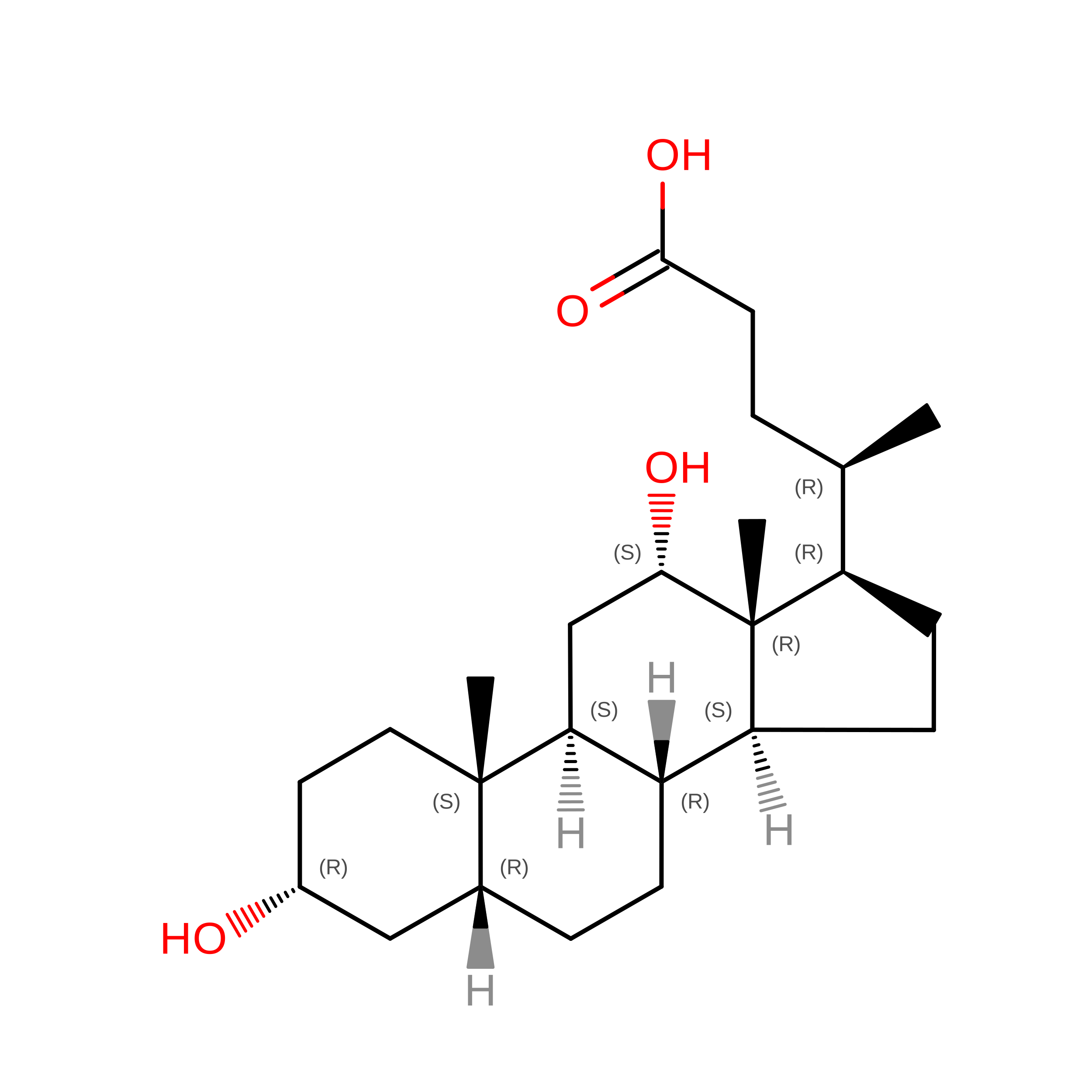
(3ALPHA,5BETA,12ALPHA)-3,12-DIHYDROXYCHOLAN-24-OIC ACID
C24 H40 O4
KXGVEGMKQFWNSR-LLQZFEROSA-N
5GP

Query on 5GP


Download:Ideal Coordinates CCD File
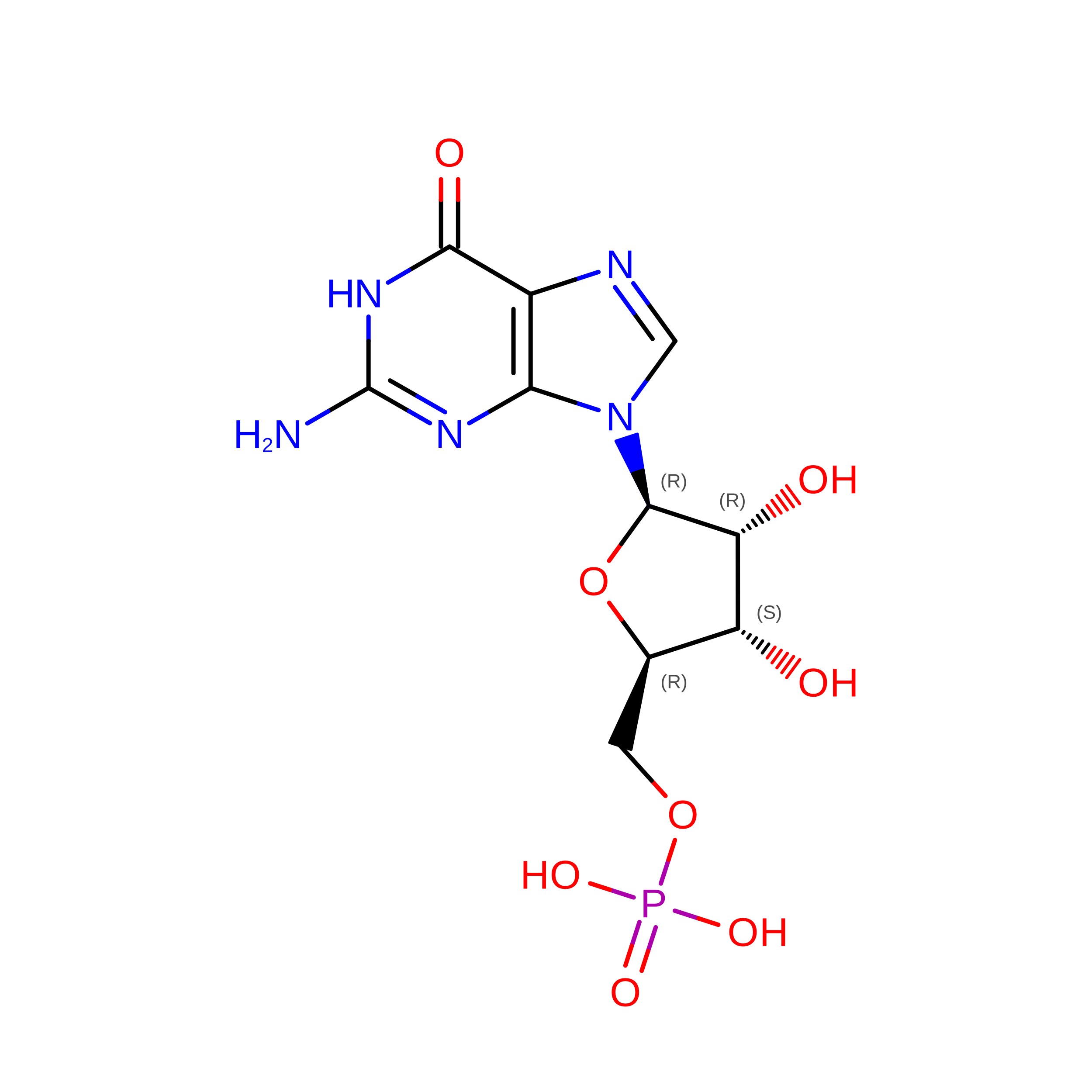
P [auth B]GUANOSINE-5'-MONOPHOSPHATE
C10 H14 N5 O8 P
RQFCJASXJCIDSX-UUOKFMHZSA-N
MMC

Query on MMC


Download:Ideal Coordinates CCD File
G [auth A]
H [auth A]
HA [auth C]
I [auth A]
IA [auth C]
G [auth A],
H [auth A],
HA [auth C],
I [auth A],
IA [auth C],
J [auth A],
JA [auth C],
KA [auth C],
LA [auth D],
MA [auth D],
NA [auth D],
OA [auth D],
Q [auth B],
R [auth B],
S [auth B],
T [auth B]
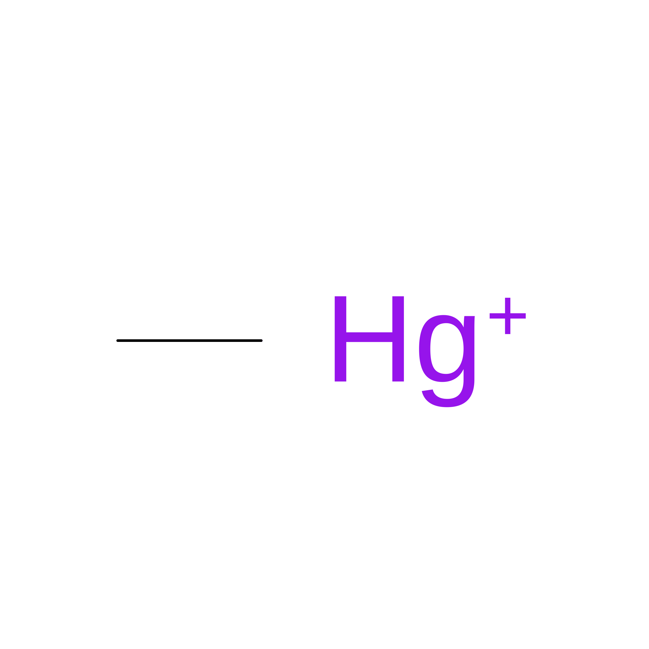
METHYL MERCURY ION
C H3 Hg
DBUXSCUEGJMZAE-UHFFFAOYSA-N
SO4

Query on SO4


Download:Ideal Coordinates CCD File
GA [auth C]
M [auth B]
N [auth B]
W [auth C]
X [auth C]
GA [auth C],
M [auth B],
N [auth B],
W [auth C],
X [auth C],
Y [auth C],
Z [auth C]
SULFATE ION
O4 S
QAOWNCQODCNURD-UHFFFAOYSA-L
MG

Query on MG


Download:Ideal Coordinates CCD File
F [auth A],
L [auth B],
V [auth C]
MAGNESIUM ION
Mg
JLVVSXFLKOJNIY-UHFFFAOYSA-N

Experimental Data & Validation

Experimental Data

  • Method: X-RAY DIFFRACTION
  • Resolution: 3.03 Å
  • R-Value Free:  0.222 (Depositor), 0.232 (DCC) 
  • R-Value Work:  0.190 (Depositor), 0.200 (DCC) 
  • R-Value Observed: 0.191 (Depositor) 
Space Group: P 31 1 2
Unit Cell:
Length ( Å )Angle ( ˚ )
a = 146.92α = 90
b = 146.92β = 90
c = 297.48γ = 120
Software Package:
Software NamePurpose
PHENIXrefinement
XDSdata reduction
XSCALEdata scaling
CNSphasing

Structure Validation

View Full Validation Report


Ligand Structure Quality Assessment 


Entry History 

Deposition Data

Revision History  (Full details and data files)

  • Version 1.0: 2012-08-22
    Type: Initial release
  • Version 1.1: 2024-11-20
    Changes: Data collection, Database references, Derived calculations, Other, Structure summary
  • Version 2.0: 2026-09-16
    Type: Remediation
    Reason: Metalloprotein remediation
    Changes: Atomic model, Data collection, Derived calculations, Non-polymer description, Polymer sequence, Structure summary