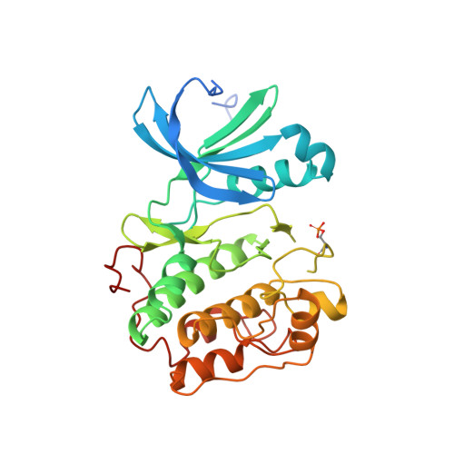
4AW1

Human PDK1 Kinase Domain in Complex with Allosteric Compound PS210 Bound to the PIF-Pocket


Experimental Data Snapshot

  • Method: X-RAY DIFFRACTION
  • Resolution: 1.68 Å
  • R-Value Free: 0.186 
  • R-Value Work: 0.164 
  • R-Value Observed: 0.165 

Starting Model: experimental
View more details

wwPDB Validation   3D Report Full Report


Ligand Structure Quality Assessment 


This is version 2.1 of the entry. See complete history


Literature

Substrate-Selective Inhibition of Protein Kinase Pdk1 by Small Compounds that Bind to the Pif-Pocket Allosteric Docking Site.

Busschots, K.Lopez-Garcia, L.A.Lammi, C.Stroba, A.Zeuzem, S.Piiper, A.Alzari, P.M.Neimanis, S.Arencibia, J.M.Engel, M.Schulze, J.O.Biondi, R.M.

(2012) Chem Biol 19: 1152

  • DOI: https://doi.org/10.1016/j.chembiol.2012.07.017
  • Primary Citation of Related Structures:  
    4AW0, 4AW1

  • PubMed Abstract: 

    The PIF-pocket of AGC protein kinases participates in the physiologic mechanism of regulation by acting as a docking site for substrates and as a switch for the transduction of the conformational changes needed for activation or inhibition. We describe the effects of compounds that bind to the PIF-pocket of PDK1. In vitro, PS210 is a potent activator of PDK1, and the crystal structure of the PDK1-ATP-PS210 complex shows that PS210 stimulates the closure of the kinase domain. However, in cells, the prodrug of PS210 (PS423) acts as a substrate-selective inhibitor of PDK1, inhibiting the phosphorylation and activation of S6K, which requires docking to the PIF-pocket, but not affecting PKB/Akt. This work describes a tool to study the dynamics of PDK1 activity and a potential approach for drug discovery.


  • Organizational Affiliation

    Research Group PhosphoSites, Department of Internal Medicine I, Universitätsklinikum Frankfurt, Theodor-Stern-Kai 7, 60590 Frankfurt, Germany.


Macromolecules
Find similar proteins by:  (by identity cutoff)  |  3D Structure
Entity ID: 1
MoleculeChains Sequence LengthOrganismDetailsImage
3-PHOSPHOINOSITIDE-DEPENDENT PROTEIN KINASE 1311Homo sapiensMutation(s): 2 
EC: 2.7.11.1
UniProt & NIH Common Fund Data Resources
Find proteins for O15530 (Homo sapiens)
Explore O15530 
Go to UniProtKB:  O15530
PHAROS:  O15530
GTEx:  ENSG00000140992 
Entity Groups  
Sequence Clusters30% Identity50% Identity70% Identity90% Identity95% Identity100% Identity
UniProt GroupO15530
Sequence Annotations
Expand
  • Reference Sequence
Small Molecules
Ligands 6 Unique
IDChains Name / Formula / InChI Key2D Diagram3D Interactions
ATP
Query on ATP

Download Ideal Coordinates CCD File 
B [auth A]ADENOSINE-5'-TRIPHOSPHATE
C10 H16 N5 O13 P3
ZKHQWZAMYRWXGA-KQYNXXCUSA-N
21O
Query on 21O

Download Ideal Coordinates CCD File 
C [auth A]{(1R)-3-oxo-1-phenyl-3-[4-(trifluoromethyl)phenyl]propyl}propanedioic acid
C19 H15 F3 O5
MLJPLHGJBUWCBA-AWEZNQCLSA-N
DMS
Query on DMS

Download Ideal Coordinates CCD File 
D [auth A],
E [auth A]
DIMETHYL SULFOXIDE
C2 H6 O S
IAZDPXIOMUYVGZ-UHFFFAOYSA-N
MN
Query on MN

Download Ideal Coordinates CCD File 
F [auth A]MANGANESE (II) ION
Mn
WAEMQWOKJMHJLA-UHFFFAOYSA-N
CL
Query on CL

Download Ideal Coordinates CCD File 
I [auth A],
J [auth A],
K [auth A]
CHLORIDE ION
Cl
VEXZGXHMUGYJMC-UHFFFAOYSA-M
NA
Query on NA

Download Ideal Coordinates CCD File 
G [auth A],
H [auth A]
SODIUM ION
Na
FKNQFGJONOIPTF-UHFFFAOYSA-N
Modified Residues  1 Unique
IDChains TypeFormula2D DiagramParent
SEP
Query on SEP
A
L-PEPTIDE LINKINGC3 H8 N O6 PSER
Binding Affinity Annotations 
IDSourceBinding Affinity
21O BindingDB:  4AW1 IC50: 3.90e+4 (nM) from 1 assay(s)
EC50: 8.58e+4 (nM) from 1 assay(s)
Experimental Data & Validation

Experimental Data

  • Method: X-RAY DIFFRACTION
  • Resolution: 1.68 Å
  • R-Value Free: 0.186 
  • R-Value Work: 0.164 
  • R-Value Observed: 0.165 
  • Space Group: C 1 2 1
Unit Cell:
Length ( Å )Angle ( ˚ )
a = 145.65α = 90
b = 42.67β = 98.52
c = 46.33γ = 90
Software Package:
Software NamePurpose
PHENIXrefinement
XDSdata reduction
XSCALEdata scaling
PHASERphasing

Structure Validation

View Full Validation Report


Ligand Structure Quality Assessment 


Entry History 

Revision History  (Full details and data files)

  • Version 1.0: 2012-10-03
    Type: Initial release
  • Version 1.1: 2012-10-10
    Changes: Database references, Derived calculations, Other, Structure summary
  • Version 2.0: 2023-12-20
    Changes: Atomic model, Data collection, Database references, Derived calculations, Other, Refinement description
  • Version 2.1: 2024-11-20
    Changes: Structure summary