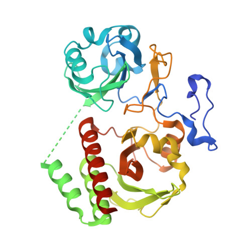
An Improved Monomeric Infrared Fluorescent Protein for Neuronal and Tumour Brain Imaging.
Yu, D., Gustafson, W.C., Han, C., Lafaye, C., Noirclerc-Savoye, M., Ge, W., Thayer, D.A., Huang, H., Kornberg, T.B., Royant, A., Jan, L.Y., Jan, Y.N., Weiss, W.A., Shu, X.(2014) Nat Commun 5: 3626
- PubMed: 24832154
- DOI: https://doi.org/10.1038/ncomms4626
- Primary Citation of Related Structures:
4CQH - PubMed Abstract:
Infrared fluorescent proteins (IFPs) are ideal for in vivo imaging, and monomeric versions of these proteins can be advantageous as protein tags or for sensor development. In contrast to GFP, which requires only molecular oxygen for chromophore maturation, phytochrome-derived IFPs incorporate biliverdin (BV) as the chromophore. However, BV varies in concentration in different cells and organisms. Here we engineered cells to express the haeme oxygenase responsible for BV biosynthesis and a brighter monomeric IFP mutant (IFP2.0). Together, these tools improve the imaging capabilities of IFP2.0 compared with monomeric IFP1.4 and dimeric iRFP. By targeting IFP2.0 to the plasma membrane, we demonstrate robust labelling of neuronal processes in Drosophila larvae. We also show that this strategy improves the sensitivity when imaging brain tumours in whole mice. Our work shows promise in the application of IFPs for protein labelling and in vivo imaging.
- 1] Department of Pharmaceutical Chemistry, University of California, San Francisco, California 94158, USA [2] Cardiovascular Research Institute, University of California, San Francisco, California 94158, USA.
Organizational Affiliation:


