4GJA

Crystal structure of renin in complex with NVP-AYL747 (compound 5)


Experimental Data Snapshot

  • Method: X-RAY DIFFRACTION
  • Resolution: 2.60 Å
  • R-Value Free: 
    0.230 (Depositor), 0.230 (DCC) 
  • R-Value Work: 
    0.185 (Depositor), 0.190 (DCC) 
  • R-Value Observed: 
    0.190 (Depositor) 

Starting Model: experimental
View more details

wwPDB Validation   3D Report Full Report


Ligand Structure Quality Assessment 

Created with Raphaël 2.3.0Worse 01 BetterLigand structure goodness of fit to experimental dataBest fitted 0M3Click on this verticalbar to view detailsBest fitted NAGClick on this verticalbar to view details

This is version 1.4 of the entry. See complete history


Literature

A novel class of oral direct Renin inhibitors: highly potent 3,5-disubstituted piperidines bearing a tricyclic p3-p1 pharmacophore.

Ostermann, N.Ruedisser, S.Ehrhardt, C.Breitenstein, W.Marzinzik, A.Jacoby, E.Vangrevelinghe, E.Ottl, J.Klumpp, M.Hartwieg, J.C.Cumin, F.Hassiepen, U.Trappe, J.Sedrani, R.Geisse, S.Gerhartz, B.Richert, P.Francotte, E.Wagner, T.Kromer, M.Kosaka, T.Webb, R.L.Rigel, D.F.Maibaum, J.Baeschlin, D.K.

(2013) J Med Chem 56: 2196-2206

  • DOI: https://doi.org/10.1021/jm301706j
  • Primary Citation of Related Structures:  
    4GJ8, 4GJ9, 4GJA, 4GJB, 4GJC, 4GJD

  • PubMed Abstract: 

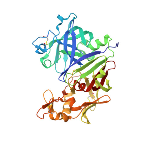
    A small library of fragments comprising putative recognition motifs for the catalytic dyad of aspartic proteases was generated by in silico similarity searches within the corporate compound deck based on rh-renin active site docking and scoring filters. Subsequent screening by NMR identified the low-affinity hits 3 and 4 as competitive active site binders, which could be shown by X-ray crystallography to bind to the hydrophobic S3-S1 pocket of rh-renin. As part of a parallel multiple hit-finding approach, the 3,5-disubstituted piperidine (rac)-5 was discovered by HTS using a enzymatic assay. X-ray crystallography demonstrated the eutomer (3S,5R)-5 to be a peptidomimetic inhibitor binding to a nonsubstrate topography of the rh-renin prime site. The design of the potent and selective (3S,5R)-12 bearing a P3(sp)-tethered tricyclic P3-P1 pharmacophore derived from 3 is described. (3S,5R)-12 showed oral bioavailability in rats and demonstrated blood pressure lowering activity in the double-transgenic rat model.


  • Organizational Affiliation

    Novartis Pharma AG, Institutes for BioMedical Research, Novartis Campus, CH-4056 Basel, Switzerland.


Macromolecules
Find similar proteins by:  (by identity cutoff)  |  3D Structure
Entity ID: 1
MoleculeChains Sequence LengthOrganismDetailsImage
Renin
A, B
340Homo sapiensMutation(s): 0 
Gene Names: REN
EC: 3.4.23.15
UniProt & NIH Common Fund Data Resources
Find proteins for P00797 (Homo sapiens)
Explore P00797 
Go to UniProtKB:  P00797
PHAROS:  P00797
GTEx:  ENSG00000143839 
Entity Groups  
Sequence Clusters30% Identity50% Identity70% Identity90% Identity95% Identity100% Identity
UniProt GroupP00797
Glycosylation
Glycosylation Sites: 1Go to GlyGen: P00797-1
Sequence Annotations
Expand
  • Reference Sequence
Small Molecules
Ligands 3 Unique
IDChains Name / Formula / InChI Key2D Diagram3D Interactions
0M3
Query on 0M3

Download Ideal Coordinates CCD File 
D [auth A],
H [auth B]
(3S,5R)-N-(2,2-diphenylethyl)-5-{[(4-methylphenyl)sulfonyl]amino}piperidine-3-carboxamide
C27 H31 N3 O3 S
TUYPQRDSHLXMFM-BJKOFHAPSA-N
NAG
Query on NAG

Download Ideal Coordinates CCD File 
C [auth A],
G [auth B]
2-acetamido-2-deoxy-beta-D-glucopyranose
C8 H15 N O6
OVRNDRQMDRJTHS-FMDGEEDCSA-N
SO4
Query on SO4

Download Ideal Coordinates CCD File 
E [auth A],
F [auth A],
I [auth B]
SULFATE ION
O4 S
QAOWNCQODCNURD-UHFFFAOYSA-L
Binding Affinity Annotations 
IDSourceBinding Affinity
0M3 BindingDB:  4GJA IC50: 1200 (nM) from 1 assay(s)
Experimental Data & Validation

Experimental Data

  • Method: X-RAY DIFFRACTION
  • Resolution: 2.60 Å
  • R-Value Free:  0.230 (Depositor), 0.230 (DCC) 
  • R-Value Work:  0.185 (Depositor), 0.190 (DCC) 
  • R-Value Observed: 0.190 (Depositor) 
Space Group: P 21 3
Unit Cell:
Length ( Å )Angle ( ˚ )
a = 141.556α = 90
b = 141.556β = 90
c = 141.556γ = 90
Software Package:
Software NamePurpose
MOLREPphasing
BUSTERrefinement
DENZOdata reduction
SCALEPACKdata scaling

Structure Validation

View Full Validation Report


Ligand Structure Quality Assessment 

Created with Raphaël 2.3.0Worse 01 BetterLigand structure goodness of fit to experimental dataBest fitted 0M3Click on this verticalbar to view detailsBest fitted NAGClick on this verticalbar to view details

Entry History 

Deposition Data

Revision History  (Full details and data files)

  • Version 1.0: 2013-02-13
    Type: Initial release
  • Version 1.1: 2013-04-10
    Changes: Database references
  • Version 1.2: 2020-07-29
    Type: Remediation
    Reason: Carbohydrate remediation
    Changes: Data collection, Derived calculations, Structure summary
  • Version 1.3: 2023-09-13
    Changes: Data collection, Database references, Refinement description, Structure summary
  • Version 1.4: 2024-11-06
    Changes: Structure summary