 Download Mendeley
Crystal structure of a GNAT superfamily phosphinothricin acetyltransferase (Pat) from Sinorhizobium meliloti 1021
Majorek, K.A., Cooper, D.R., Ahmed, M., Stead, M., Hillerich, B., Seidel, R., Bonanno, J.B., Almo, S.C., Minor, W.To be published.
Experimental Data Snapshot
wwPDB Validation 3D Report Full Report

Entity ID: 1 | |||||
|---|---|---|---|---|---|
| Molecule | Chains | Sequence Length | Organism | Details | Image |
| Acetyltransferase | 186 | Sinorhizobium meliloti 1021 | Mutation(s): 0 Gene Names: R03223, SMc03840 EC: 2.3.1 |  | |
UniProt | |||||
Entity Groups | |||||
| Sequence Clusters | 30% Identity50% Identity70% Identity90% Identity95% Identity100% Identity | ||||
| UniProt Group | Q92L60 | ||||
Sequence AnnotationsExpand | |||||
Reference Sequence | |||||
| Ligands 4 Unique | |||||
|---|---|---|---|---|---|
| ID | Chains | Name / Formula / InChI Key | 2D Diagram | 3D Interactions | |
| 2PE Download:Ideal Coordinates CCD File | E [auth A] | NONAETHYLENE GLYCOL C18 H38 O10 YZUUTMGDONTGTN-UHFFFAOYSA-N | 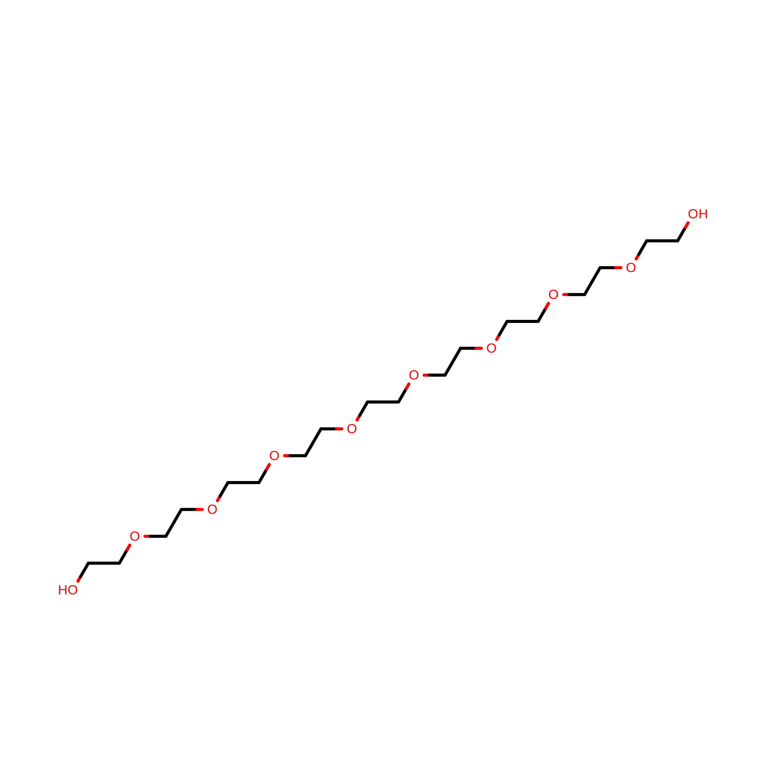 | ||
| FLC Download:Ideal Coordinates CCD File | D [auth A], H [auth B] | CITRATE ANION C6 H5 O7 KRKNYBCHXYNGOX-UHFFFAOYSA-K | 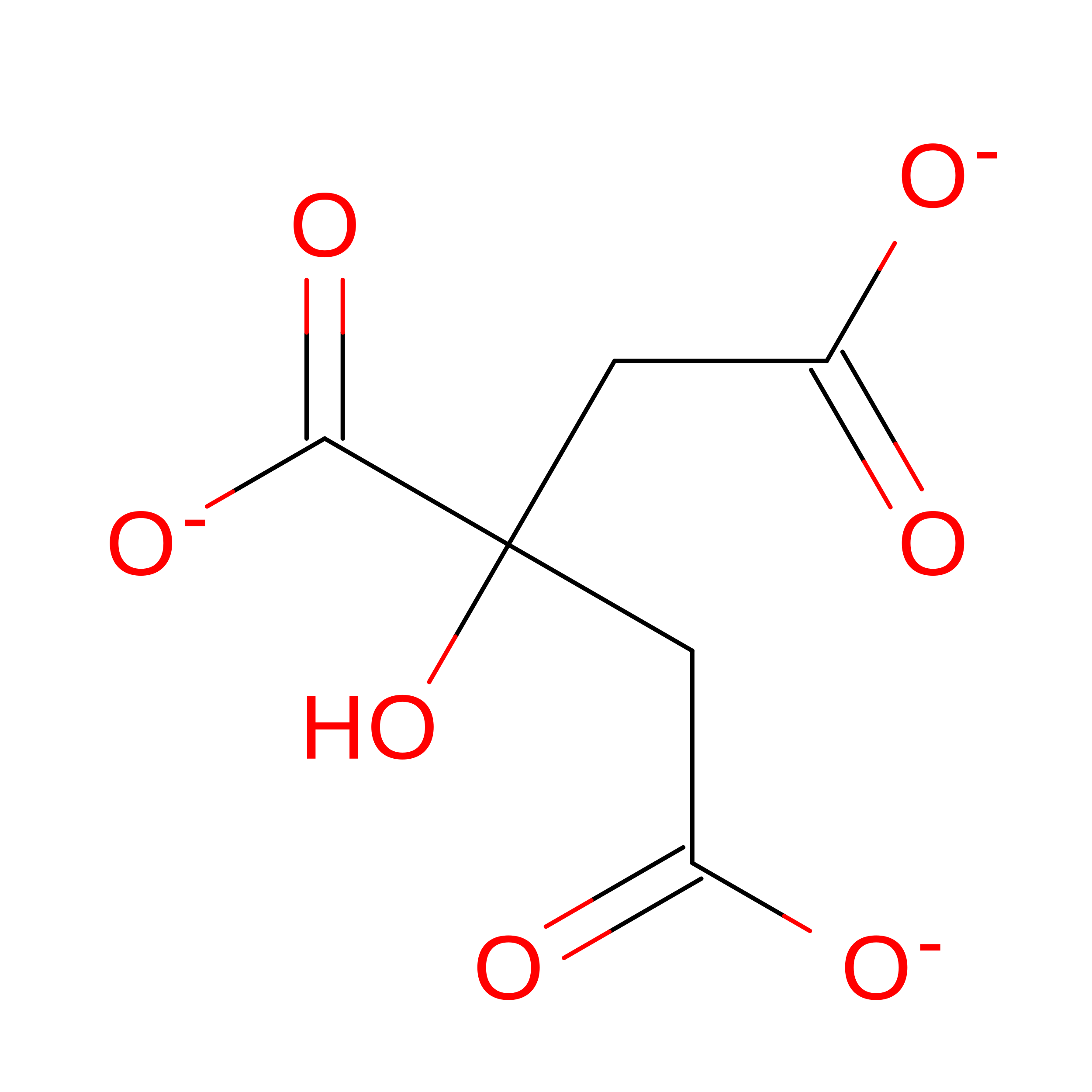 | ||
| PEG Download:Ideal Coordinates CCD File | F [auth B] | DI(HYDROXYETHYL)ETHER C4 H10 O3 MTHSVFCYNBDYFN-UHFFFAOYSA-N |  | ||
| EDO Download:Ideal Coordinates CCD File | C [auth A], G [auth B] | 1,2-ETHANEDIOL C2 H6 O2 LYCAIKOWRPUZTN-UHFFFAOYSA-N |  | ||
| Modified Residues 1 Unique | |||||
|---|---|---|---|---|---|
| ID | Chains | Modified Residue | Modification Type | Formula | 2D Diagram |
| MSE Query on MSE | MET | Selenomethionine | C5 H11 N O2 Se |  | |
| Length ( Å ) | Angle ( ˚ ) |
|---|---|
| a = 51.461 | α = 90 |
| b = 80.481 | β = 90 |
| c = 83.611 | γ = 90 |
| Software Name | Purpose |
|---|---|
| HKL-3000 | data collection |
| HKL-3000 | phasing |
| SHELXD | phasing |
| SHELXE | model building |
| MLPHARE | phasing |
| DM | model building |
| ARP/wARP | model building |
| REFMAC | refinement |
| Coot | model building |
| HKL-3000 | data reduction |
| HKL-3000 | data scaling |
| DM | phasing |
