4NXO | pdb_00004nxo

Crystal Structure of Insulin Degrading Enzyme in complex with BDM44768


Experimental Data Snapshot

  • Method: X-RAY DIFFRACTION
  • Resolution: 2.73 Å
  • R-Value Free: 
    0.219 (Depositor), 0.220 (DCC) 
  • R-Value Work: 
    0.170 (Depositor), 0.170 (DCC) 
  • R-Value Observed: 
    0.171 (Depositor) 

Starting Model: experimental
View more details

wwPDB Validation   3D Report Full Report


Ligand Structure Quality Assessment 

Created with Raphaël 2.3.0Worse 01 BetterLigand structure goodness of fit to experimental dataBest fitted 2H7Click on this verticalbar to view details

This is version 1.3 of the entry. See complete history


Literature

Catalytic site inhibition of insulin-degrading enzyme by a small molecule induces glucose intolerance in mice.

Deprez-Poulain, R.Hennuyer, N.Bosc, D.Liang, W.G.Enee, E.Marechal, X.Charton, J.Totobenazara, J.Berte, G.Jahklal, J.Verdelet, T.Dumont, J.Dassonneville, S.Woitrain, E.Gauriot, M.Paquet, C.Duplan, I.Hermant, P.Cantrelle, F.X.Sevin, E.Culot, M.Landry, V.Herledan, A.Piveteau, C.Lippens, G.Leroux, F.Tang, W.J.van Endert, P.Staels, B.Deprez, B.

(2015) Nat Commun 6: 8250-8250

  • DOI: https://doi.org/10.1038/ncomms9250
  • Primary Citation of Related Structures:  
    4IFH, 4NXO, 4RE9

  • PubMed Abstract: 

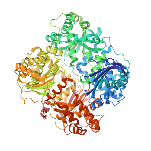
    Insulin-degrading enzyme (IDE) is a protease that cleaves insulin and other bioactive peptides such as amyloid-β. Knockout and genetic studies have linked IDE to Alzheimer's disease and type-2 diabetes. As the major insulin-degrading protease, IDE is a candidate drug target in diabetes. Here we have used kinetic target-guided synthesis to design the first catalytic site inhibitor of IDE suitable for in vivo studies (BDM44768). Crystallographic and small angle X-ray scattering analyses show that it locks IDE in a closed conformation. Among a panel of metalloproteases, BDM44768 selectively inhibits IDE. Acute treatment of mice with BDM44768 increases insulin signalling and surprisingly impairs glucose tolerance in an IDE-dependent manner. These results confirm that IDE is involved in pathways that modulate short-term glucose homeostasis, but casts doubt on the general usefulness of the inhibition of IDE catalytic activity to treat diabetes.


  • Organizational Affiliation

    Institut Pasteur de Lille, Lille F-59000, France.


Macromolecules
Find similar proteins by:  (by identity cutoff)  |  3D Structure
Entity ID: 1
MoleculeChains Sequence LengthOrganismDetailsImage
Insulin-degrading enzyme
A, B
990Homo sapiensMutation(s): 14 
Gene Names: IDE
EC: 3.4.24.56
UniProt & NIH Common Fund Data Resources
Find proteins for P14735 (Homo sapiens)
Explore P14735 
Go to UniProtKB:  P14735
PHAROS:  P14735
GTEx:  ENSG00000119912 
Entity Groups  
Sequence Clusters30% Identity50% Identity70% Identity90% Identity95% Identity100% Identity
UniProt GroupP14735
Sequence Annotations
Expand
  • Reference Sequence
Small Molecules
Ligands 7 Unique
IDChains Name / Formula / InChI Key2D Diagram3D Interactions
2H7
Query on 2H7

Download Ideal Coordinates CCD File 
D [auth A],
M [auth B]
4-fluoro-N-({1-[(2R)-4-(hydroxyamino)-1-(naphthalen-2-yl)-4-oxobutan-2-yl]-1H-1,2,3-triazol-4-yl}methyl)benzamide
C24 H22 F N5 O3
RASCFMYPAZDCKQ-JOCHJYFZSA-N
EPE
Query on EPE

Download Ideal Coordinates CCD File 
K [auth A],
U [auth B]
4-(2-HYDROXYETHYL)-1-PIPERAZINE ETHANESULFONIC ACID
C8 H18 N2 O4 S
JKMHFZQWWAIEOD-UHFFFAOYSA-N
1PE
Query on 1PE

Download Ideal Coordinates CCD File 
E [auth A],
N [auth B]
PENTAETHYLENE GLYCOL
C10 H22 O6
JLFNLZLINWHATN-UHFFFAOYSA-N
PEG
Query on PEG

Download Ideal Coordinates CCD File 
F [auth A]
G [auth A]
H [auth A]
O [auth B]
P [auth B]
DI(HYDROXYETHYL)ETHER
C4 H10 O3
MTHSVFCYNBDYFN-UHFFFAOYSA-N
DIO
Query on DIO

Download Ideal Coordinates CCD File 
V [auth B]1,4-DIETHYLENE DIOXIDE
C4 H8 O2
RYHBNJHYFVUHQT-UHFFFAOYSA-N
ZN
Query on ZN

Download Ideal Coordinates CCD File 
C [auth A],
L [auth B]
ZINC ION
Zn
PTFCDOFLOPIGGS-UHFFFAOYSA-N
EDO
Query on EDO

Download Ideal Coordinates CCD File 
I [auth A],
J [auth A],
R [auth B],
S [auth B]
1,2-ETHANEDIOL
C2 H6 O2
LYCAIKOWRPUZTN-UHFFFAOYSA-N
Binding Affinity Annotations 
IDSourceBinding Affinity
2H7 BindingDB:  4NXO IC50: min: 56, max: 330 (nM) from 3 assay(s)
Experimental Data & Validation

Experimental Data

  • Method: X-RAY DIFFRACTION
  • Resolution: 2.73 Å
  • R-Value Free:  0.219 (Depositor), 0.220 (DCC) 
  • R-Value Work:  0.170 (Depositor), 0.170 (DCC) 
  • R-Value Observed: 0.171 (Depositor) 
Space Group: P 65
Unit Cell:
Length ( Å )Angle ( ˚ )
a = 262.909α = 90
b = 262.909β = 90
c = 86.337γ = 120
Software Package:
Software NamePurpose
HKL-3000data collection
PHENIXmodel building
PHENIXrefinement
HKL-3000data reduction
HKL-3000data scaling
PHASERphasing

Structure Validation

View Full Validation Report


Ligand Structure Quality Assessment 

Created with Raphaël 2.3.0Worse 01 BetterLigand structure goodness of fit to experimental dataBest fitted 2H7Click on this verticalbar to view details

Entry History 

Deposition Data

Revision History  (Full details and data files)

  • Version 1.0: 2015-10-07
    Type: Initial release
  • Version 1.1: 2015-10-14
    Changes: Data collection
  • Version 1.2: 2015-10-28
    Changes: Database references
  • Version 1.3: 2023-09-20
    Changes: Data collection, Database references, Derived calculations, Refinement description