4PJZ

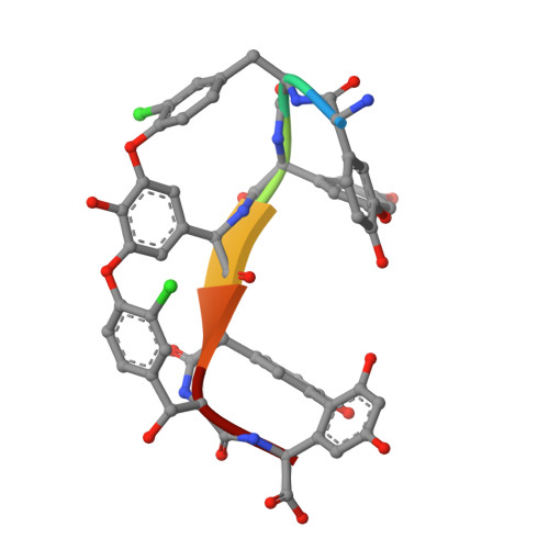
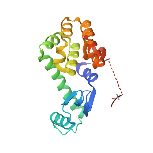
CRYSTAL STRUCTURE OF T4 LYSOZYME-GSS-PEPTIDE IN COMPLEX WITH TEICOPLANIN-A2-2


Experimental Data Snapshot

  • Method: X-RAY DIFFRACTION
  • Resolution: 1.87 Å
  • R-Value Free: 0.228 
  • R-Value Work: 0.194 
  • R-Value Observed: 0.196 

wwPDB Validation   3D Report Full Report


Ligand Structure Quality Assessment 


This is version 2.2 of the entry. See complete history


Literature

X-ray Crystal Structure of Teicoplanin A2-2 Bound to a Catalytic Peptide Sequence via the Carrier Protein Strategy.

Han, S.Le, B.V.Hajare, H.S.Baxter, R.H.Miller, S.J.

(2014) J Org Chem 79: 8550-8556

  • DOI: https://doi.org/10.1021/jo501625f
  • Primary Citation of Related Structures:  
    4PJZ, 4PK0

  • PubMed Abstract: 

    We report the X-ray crystal structure of a site-selective peptide catalyst moiety and teicoplanin A2-2 complex. The expressed protein ligation technique was used to couple T4 lysozyme (T4L) and a synthetic peptide catalyst responsible for the selective phosphorylation of the N-acetylglucosamine sugar in a teicoplanin A2-2 derivative. The T4L-Pmh-dPro-Aib-dAla-dAla construct was crystallized in the presence of teicoplanin A2-2. The resulting 2.3 Å resolution protein-peptide-teicoplanin complex crystal structure revealed that the nucleophilic nitrogen of N-methylimidazole in the Pmh residue is in closer proximity (7.6 Å) to the N-acetylglucosamine than the two other sugar rings present in teicoplanin (9.3 and 20.3 Å, respectively). This molecular arrangement is consistent with the observed selectivity afforded by the peptide-based catalyst when it is applied to a site-selective phosphorylation reaction involving a teicoplanin A2-2 derivative.


  • Organizational Affiliation

    Department of Chemistry, Yale University , New Haven, Connecticut 06511, United States.


Macromolecules

Find similar proteins by:  Sequence   |   3D Structure  

Entity ID: 1
MoleculeChains Sequence LengthOrganismDetailsImage
TEICOPLANIN-A2-2A [auth B]7Actinoplanes teichomyceticusMutation(s): 0 
Sequence Annotations
Expand
  • Reference Sequence
Find similar proteins by:  (by identity cutoff)  |  3D Structure
Entity ID: 2
MoleculeChains Sequence LengthOrganismDetailsImage
LysozymeB [auth A]174Tequatrovirus T4Mutation(s): 2 
Gene Names: eT4Tp126
EC: 3.2.1.17
UniProt
Find proteins for D9IEF7 (Enterobacteria phage T4)
Explore D9IEF7 
Go to UniProtKB:  D9IEF7
Entity Groups  
Sequence Clusters30% Identity50% Identity70% Identity90% Identity95% Identity100% Identity
UniProt GroupD9IEF7
Sequence Annotations
Expand
  • Reference Sequence
Small Molecules
Ligands 6 Unique
IDChains Name / Formula / InChI Key2D Diagram3D Interactions
NAG
Query on NAG

Download Ideal Coordinates CCD File 
E [auth B]2-acetamido-2-deoxy-beta-D-glucopyranose
C8 H15 N O6
OVRNDRQMDRJTHS-FMDGEEDCSA-N
MAN
Query on MAN

Download Ideal Coordinates CCD File 
F [auth B]alpha-D-mannopyranose
C6 H12 O6
WQZGKKKJIJFFOK-PQMKYFCFSA-N
GCS
Query on GCS

Download Ideal Coordinates CCD File 
C [auth B]2-amino-2-deoxy-beta-D-glucopyranose
C6 H13 N O5
MSWZFWKMSRAUBD-QZABAPFNSA-N
T55
Query on T55

Download Ideal Coordinates CCD File 
D [auth B]8-METHYLNONANOIC ACID
C10 H20 O2
OAOABCKPVCUNKO-UHFFFAOYSA-N
PO4
Query on PO4

Download Ideal Coordinates CCD File 
G [auth B],
I [auth A]
PHOSPHATE ION
O4 P
NBIIXXVUZAFLBC-UHFFFAOYSA-K
GOL
Query on GOL

Download Ideal Coordinates CCD File 
H [auth B],
J [auth A]
GLYCEROL
C3 H8 O3
PEDCQBHIVMGVHV-UHFFFAOYSA-N
Modified Residues  5 Unique
IDChains TypeFormula2D DiagramParent
3MY
Query on 3MY
A [auth B]D-PEPTIDE LINKINGC9 H10 Cl N O3TYR
OMY
Query on OMY
A [auth B]L-PEPTIDE LINKINGC9 H10 Cl N O4TYR
AIB
Query on AIB
B [auth A]L-PEPTIDE LINKINGC4 H9 N O2ALA
CCS
Query on CCS
B [auth A]L-PEPTIDE LINKINGC5 H9 N O4 SCYS
MHS
Query on MHS
B [auth A]L-PEPTIDE LINKINGC7 H11 N3 O2HIS
Biologically Interesting Molecules (External Reference) 1 Unique
Experimental Data & Validation

Experimental Data

  • Method: X-RAY DIFFRACTION
  • Resolution: 1.87 Å
  • R-Value Free: 0.228 
  • R-Value Work: 0.194 
  • R-Value Observed: 0.196 
  • Space Group: I 2 2 2
Unit Cell:
Length ( Å )Angle ( ˚ )
a = 58.116α = 90
b = 63.569β = 90
c = 138.884γ = 90
Software Package:
Software NamePurpose
REFMACrefinement

Structure Validation

View Full Validation Report


Ligand Structure Quality Assessment 


Entry History & Funding Information

Deposition Data


Funding OrganizationLocationGrant Number
National Institutes of Health/National Institute of General Medical Sciences (NIH/NIGMS)United StatesGM-068649

Revision History  (Full details and data files)

  • Version 1.0: 2014-09-10
    Type: Initial release
  • Version 1.1: 2014-10-01
    Changes: Database references
  • Version 1.2: 2017-09-20
    Changes: Author supporting evidence, Database references, Derived calculations, Other, Source and taxonomy, Structure summary
  • Version 2.0: 2019-12-25
    Changes: Author supporting evidence, Polymer sequence
  • Version 2.1: 2020-07-29
    Type: Remediation
    Reason: Carbohydrate remediation
    Changes: Data collection, Derived calculations, Refinement description, Structure summary
  • Version 2.2: 2023-12-27
    Changes: Data collection, Database references, Derived calculations, Structure summary