4QJK

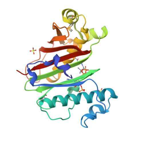
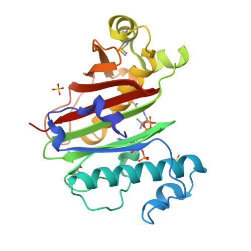
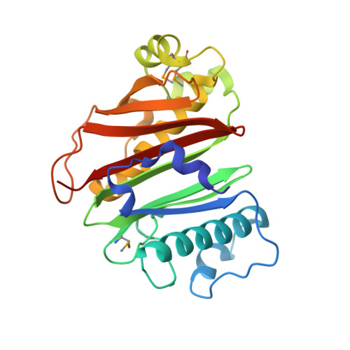
Crystal structure of M. tuberculosis phosphopantetheinyl transferase PptT


Experimental Data Snapshot

  • Method: X-RAY DIFFRACTION
  • Resolution: 1.59 Å
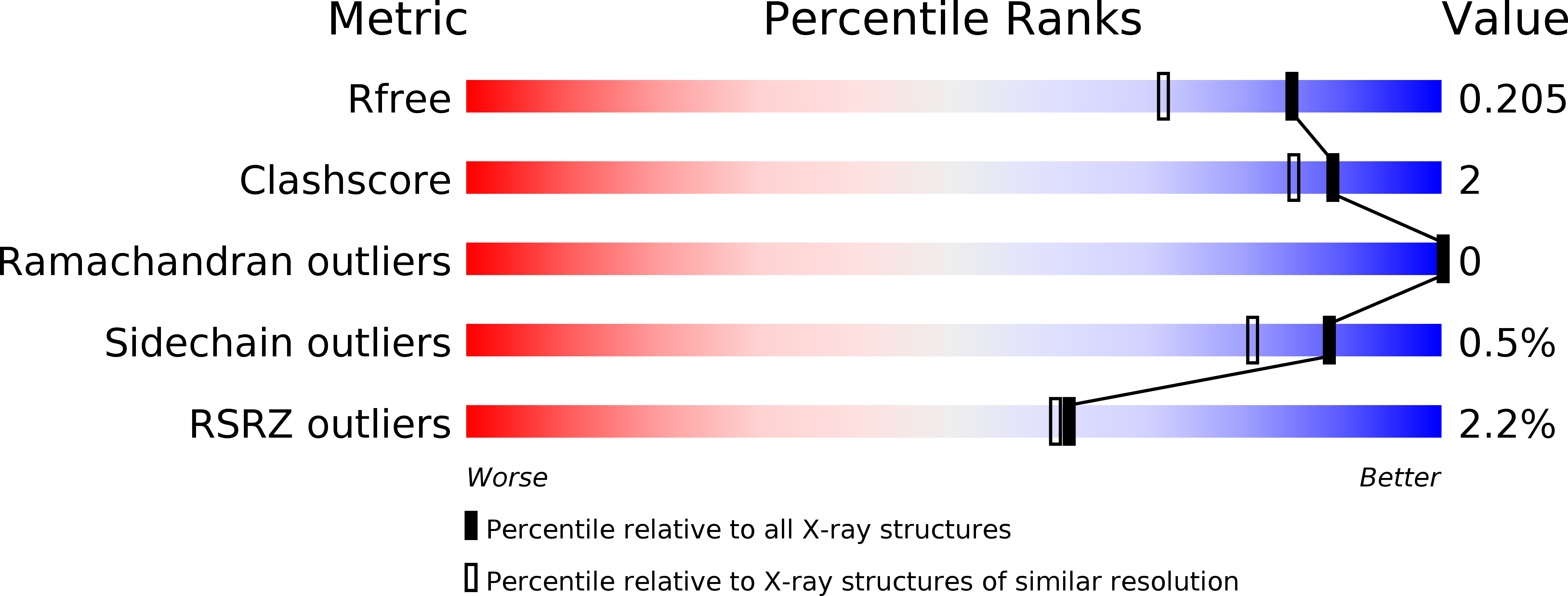
  • R-Value Free: 0.204 
  • R-Value Work: 0.170 
  • R-Value Observed: 0.171 

wwPDB Validation   3D Report Full Report


Ligand Structure Quality Assessment 


This is version 1.3 of the entry. See complete history


Literature

Structure, biochemistry, and inhibition of essential 4'-phosphopantetheinyl transferases from two species of mycobacteria.

Vickery, C.R.Kosa, N.M.Casavant, E.P.Duan, S.Noel, J.P.Burkart, M.D.

(2014) ACS Chem Biol 9: 1939-1944

  • DOI: https://doi.org/10.1021/cb500263p
  • Primary Citation of Related Structures:  
    4QJK, 4QJL

  • PubMed Abstract: 

    4'-Phosphopantetheinyl transferases (PPTase) post-translationally modify carrier proteins with a phosphopantetheine moiety, an essential reaction in all three domains of life. In the bacterial genus Mycobacteria, the Sfp-type PPTase activates pathways necessary for the biosynthesis of cell wall components and small molecule virulence factors. We solved the X-ray crystal structures and biochemically characterized the Sfp-type PPTases from two of the most prevalent Mycobacterial pathogens, PptT of M. tuberculosis and MuPPT of M. ulcerans. Structural analyses reveal significant differences in cofactor binding and active site composition when compared to previously characterized Sfp-type PPTases. Functional analyses including the efficacy of Sfp-type PPTase-specific inhibitors also suggest that the Mycobacterial Sfp-type PPTases can serve as therapeutic targets against Mycobacterial infections.


  • Organizational Affiliation

    Department of Chemistry and Biochemistry, University of California, San Diego , 9500 Gilman Drive, La Jolla, California 92093-0358, United States.


Macromolecules
Find similar proteins by:  (by identity cutoff)  |  3D Structure
Entity ID: 1
MoleculeChains Sequence LengthOrganismDetailsImage
Phosphopantetheinyl transferase PptT235Mycobacterium tuberculosis H37RvMutation(s): 0 
Gene Names: NP_217310pptTRv2794cRVBD_2794c
EC: 2.7.8.7
UniProt
Find proteins for O33336 (Mycobacterium tuberculosis (strain ATCC 25618 / H37Rv))
Explore O33336 
Go to UniProtKB:  O33336
Entity Groups  
Sequence Clusters30% Identity50% Identity70% Identity90% Identity95% Identity100% Identity
UniProt GroupO33336
Sequence Annotations
Expand
  • Reference Sequence
Small Molecules
Modified Residues  1 Unique
IDChains TypeFormula2D DiagramParent
MSE
Query on MSE
A
L-PEPTIDE LINKINGC5 H11 N O2 SeMET
Experimental Data & Validation

Experimental Data

  • Method: X-RAY DIFFRACTION
  • Resolution: 1.59 Å
  • R-Value Free: 0.204 
  • R-Value Work: 0.170 
  • R-Value Observed: 0.171 
  • Space Group: C 2 2 21
Unit Cell:
Length ( Å )Angle ( ˚ )
a = 99.86α = 90
b = 121.505β = 90
c = 48.78γ = 90
Software Package:
Software NamePurpose
MOSFLMdata reduction
SCALAdata scaling
PHENIXrefinement
PDB_EXTRACTdata extraction
BOSdata collection
AutoSolphasing

Structure Validation

View Full Validation Report


Ligand Structure Quality Assessment 


Entry History 

Deposition Data

Revision History  (Full details and data files)

  • Version 1.0: 2014-07-16
    Type: Initial release
  • Version 1.1: 2014-10-01
    Changes: Database references
  • Version 1.2: 2017-11-22
    Changes: Refinement description
  • Version 1.3: 2024-10-30
    Changes: Data collection, Database references, Derived calculations, Structure summary