4W1X | pdb_00004w1x

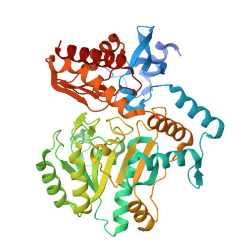
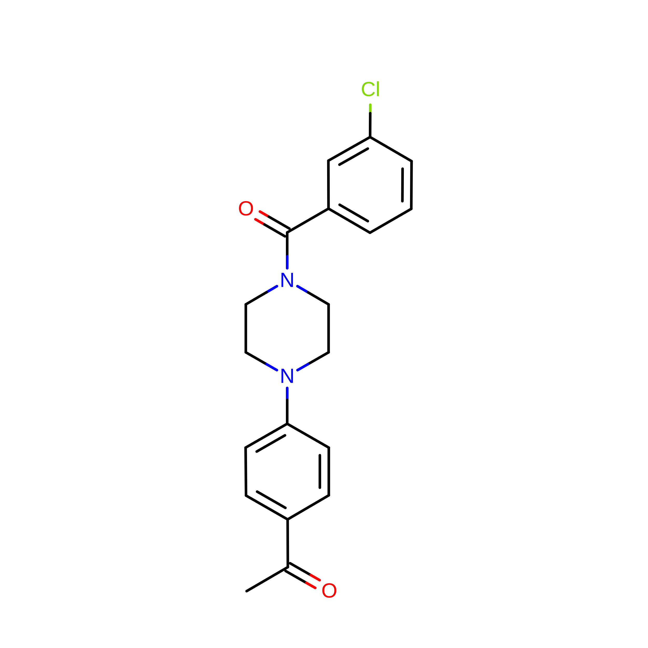
Crystal structure of 7,8-diaminopelargonic acid synthase (BioA) from Mycobacterium tuberculosis, complexed with 1-(4-(4-(3-chlorobenzoyl)piperazin-1-yl)phenyl)ethanone


Experimental Data Snapshot

  • Method: X-RAY DIFFRACTION
  • Resolution: 1.80 Å
  • R-Value Free: 
    0.227 (Depositor), 0.223 (DCC) 
  • R-Value Work: 
    0.198 (Depositor), 0.193 (DCC) 
  • R-Value Observed: 
    0.199 (Depositor) 

wwPDB Validation 3D Report Full Report

Validation slider image for 4W1X

Ligand Structure Quality Assessment 


This is version 2.1 of the entry. See complete history. 

Literature

Target-Based Identification of Whole-Cell Active Inhibitors of Biotin Biosynthesis in Mycobacterium tuberculosis.

Park, S.W., Casalena, D.E., Wilson, D.J., Dai, R., Nag, P.P., Liu, F., Boyce, J.P., Bittker, J.A., Schreiber, S.L., Finzel, B.C., Schnappinger, D., Aldrich, C.C.

(2015) Chem Biol 22: 76-86

  • DOI: https://doi.org/10.1016/j.chembiol.2014.11.012
  • Primary Citation Related Structures: 
    4W1V, 4W1W, 4W1X

  • PubMed Abstract: 

    Biotin biosynthesis is essential for survival and persistence of Mycobacterium tuberculosis (Mtb) in vivo. The aminotransferase BioA, which catalyzes the antepenultimate step in the biotin pathway, has been established as a promising target due to its vulnerability to chemical inhibition. We performed high-throughput screening (HTS) employing a fluorescence displacement assay and identified a diverse set of potent inhibitors including many diversity-oriented synthesis (DOS) scaffolds. To efficiently select only hits targeting biotin biosynthesis, we then deployed a whole-cell counterscreen in biotin-free and biotin-containing medium against wild-type Mtb and in parallel with isogenic bioA Mtb strains that possess differential levels of BioA expression. This counterscreen proved crucial to filter out compounds whose whole-cell activity was off target as well as identify hits with weak, but measurable whole-cell activity in BioA-depleted strains. Several of the most promising hits were cocrystallized with BioA to provide a framework for future structure-based drug design efforts.


  • Organizational Affiliation: 
    • Department of Microbiology and Immunology, Weill Cornell Medical College, New York, NY 10065, USA.

Macromolecule Content 

  • Total Structure Weight: 98.55 kDa 
  • Atom Count: 7,133 
  • Modeled Residue Count: 858 
  • Deposited Residue Count: 914 
  • Unique protein chains: 1

Macromolecules

Find similar proteins by:|  3D Structure
Entity ID: 1
MoleculeChains  Sequence LengthOrganismDetailsImage
Adenosylmethionine-8-amino-7-oxononanoate aminotransferase
A, B
457Mycobacterium tuberculosis CDC1551Mutation(s): 0 
Gene Names: bioA, MT1619
EC: 2.6.1.62
UniProt
Find proteins for P9WQ80 (Mycobacterium tuberculosis (strain CDC 1551 / Oshkosh))
Explore P9WQ80 
Go to UniProtKB:  P9WQ80
Entity Groups
Sequence Clusters30% Identity50% Identity70% Identity90% Identity95% Identity100% Identity
UniProt GroupP9WQ80
Sequence Annotations
Expand
Reference Sequence

Small Molecules

Ligands 4 Unique
IDChains Name / Formula / InChI Key2D Diagram3D Interactions
3G9

Query on 3G9


Download:Ideal Coordinates CCD File
G [auth B],
H [auth B]
1-{4-[4-(3-chlorobenzoyl)piperazin-1-yl]phenyl}ethanone
C19 H19 Cl N2 O2
RCHDWCNBGCYJNK-UHFFFAOYSA-N
PLP

Query on PLP


Download:Ideal Coordinates CCD File
C [auth A],
J [auth B]
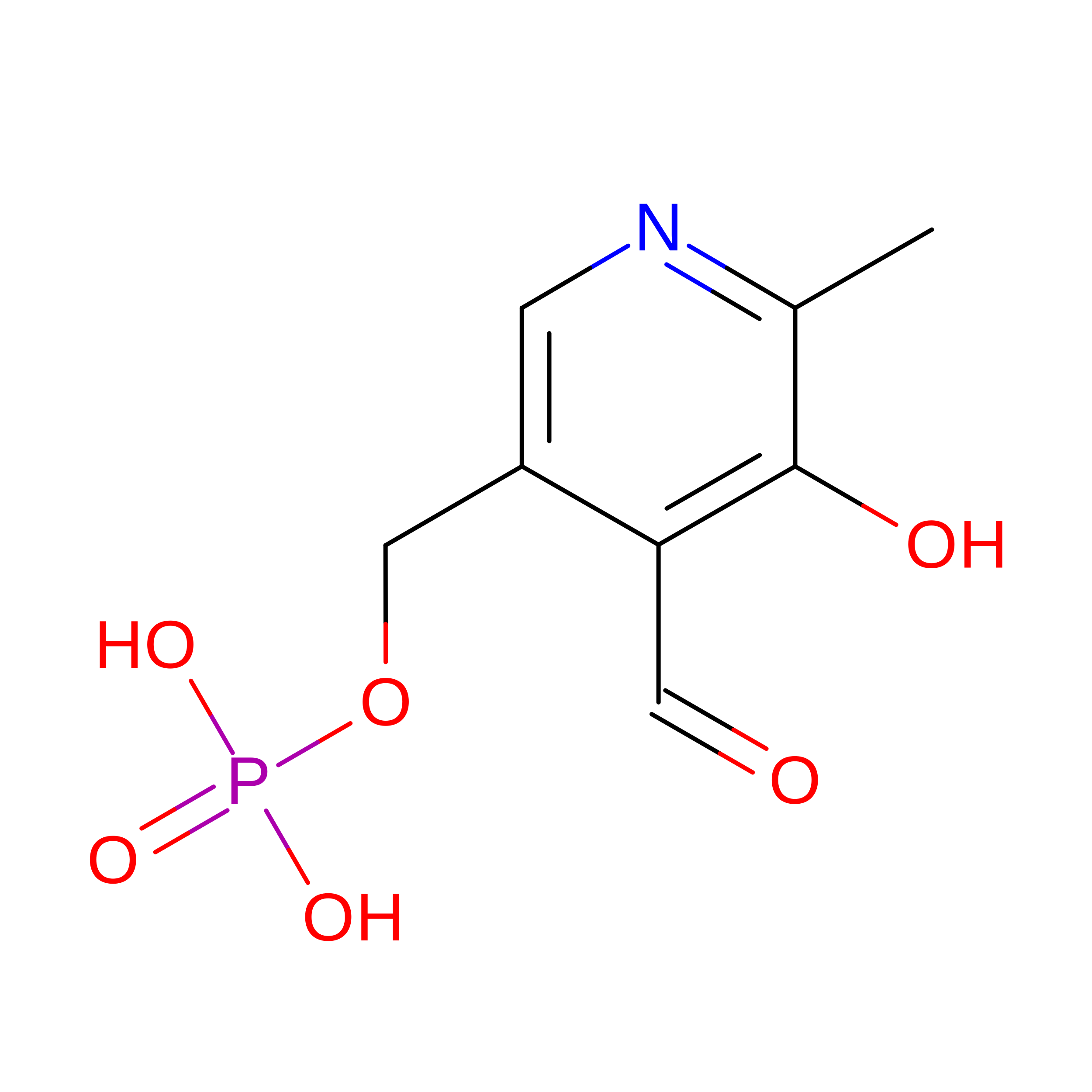
PYRIDOXAL-5'-PHOSPHATE
C8 H10 N O6 P
NGVDGCNFYWLIFO-UHFFFAOYSA-N
PEG

Query on PEG


Download:Ideal Coordinates CCD File
E [auth A]DI(HYDROXYETHYL)ETHER
C4 H10 O3
MTHSVFCYNBDYFN-UHFFFAOYSA-N
EDO

Query on EDO


Download:Ideal Coordinates CCD File
D [auth A],
F [auth A],
I [auth B]
1,2-ETHANEDIOL
C2 H6 O2
LYCAIKOWRPUZTN-UHFFFAOYSA-N

Experimental Data & Validation

Experimental Data

  • Method: X-RAY DIFFRACTION
  • Resolution: 1.80 Å
  • R-Value Free:  0.227 (Depositor), 0.223 (DCC) 
  • R-Value Work:  0.198 (Depositor), 0.193 (DCC) 
  • R-Value Observed: 0.199 (Depositor) 
Space Group: P 21 21 21
Unit Cell:
Length ( Å )Angle ( ˚ )
a = 62α = 90
b = 66β = 90
c = 204γ = 90
Software Package:
Software NamePurpose
d*TREKdata reduction
REFMACrefinement
PDB_EXTRACTdata extraction
d*TREKdata scaling
d*TREKdata scaling

Structure Validation

View Full Validation Report


Ligand Structure Quality Assessment 


Entry History 

Deposition Data

Revision History  (Full details and data files)

  • Version 1.0: 2015-02-04
    Type: Initial release
  • Version 2.0: 2017-11-22
    Changes: Atomic model, Database references, Derived calculations, Other, Refinement description, Source and taxonomy
  • Version 2.1: 2023-12-27
    Changes: Data collection, Database references, Derived calculations, Refinement description