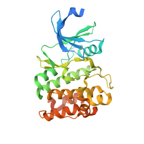
RCSB PDB - 4X7Q: PIM2 kinase in complex with Compound 1s

 4X7Q

PIM2 kinase in complex with Compound 1s


Experimental Data Snapshot

  • Method: X-RAY DIFFRACTION
  • Resolution: 2.33 Å
  • R-Value Free: 0.258 
  • R-Value Work: 0.196 
  • R-Value Observed: 0.199 

Starting Model: experimental
View more details

wwPDB Validation   3D Report Full Report


Ligand Structure Quality Assessment 

Created with Raphaël 2.3.0Worse 01 BetterLigand structure goodness of fit to experimental dataBest fitted 3YRClick on this verticalbar to view details

This is version 1.2 of the entry. See complete history


Literature

Structure-based design of low-nanomolar PIM kinase inhibitors.

Ishchenko, A.Zhang, L.Le Brazidec, J.Y.Fan, J.Chong, J.H.Hingway, A.Raditsis, A.Singh, L.Elenbaas, B.Hong, V.S.Marcotte, D.Silvian, L.Enyedy, I.Chao, J.

(2015) Bioorg Med Chem Lett 25: 474-480

  • DOI: https://doi.org/10.1016/j.bmcl.2014.12.041
  • Primary Citation of Related Structures:  
    4X7Q, 4XHK

  • PubMed Abstract: 

    PIM kinases are implicated in variety of cancers by promoting cell survival and proliferation and are targets of interest for therapeutic intervention. We have identified a low-nanomolar pan-PIM inhibitor (PIM1/2/3 potency 5:14:2nM) using structure based modeling. The crystal structure of this compound with PIM1 confirmed the predicted binding mode and protein-ligand interactions except those in the acidic ribose pocket. We show the SAR suggesting the importance of having a hydrogen bond donor in this pocket for inhibiting PIM2; however, this interaction is not important for inhibiting PIM1 or PIM3. In addition, we report the discovery of a new class of PIM inhibitors by using computational de novo design tool implemented in MOE software (Chemical Computing Group). These inhibitors have a different interaction profile.


  • Organizational Affiliation

    Biogen Idec, 14 Cambridge Center, Cambridge, MA 02142, USA. Electronic address: al_ishchenko@yahoo.com.


Macromolecules
Find similar proteins by:  (by identity cutoff)  |  3D Structure
Entity ID: 1
MoleculeChains Sequence LengthOrganismDetailsImage
Serine/threonine-protein kinase pim-2A [auth B],
B [auth A]
312Homo sapiensMutation(s): 0 
Gene Names: PIM2
EC: 2.7.11.1
UniProt & NIH Common Fund Data Resources
Find proteins for Q9P1W9 (Homo sapiens)
Explore Q9P1W9 
Go to UniProtKB:  Q9P1W9
PHAROS:  Q9P1W9
GTEx:  ENSG00000102096 
Entity Groups  
Sequence Clusters30% Identity50% Identity70% Identity90% Identity95% Identity100% Identity
UniProt GroupQ9P1W9
Sequence Annotations
Expand
  • Reference Sequence
Small Molecules
Binding Affinity Annotations 
IDSourceBinding Affinity
3YR BindingDB:  4X7Q IC50: 14 (nM) from 1 assay(s)
Experimental Data & Validation

Experimental Data

  • Method: X-RAY DIFFRACTION
  • Resolution: 2.33 Å
  • R-Value Free: 0.258 
  • R-Value Work: 0.196 
  • R-Value Observed: 0.199 
  • Space Group: H 3
Unit Cell:
Length ( Å )Angle ( ˚ )
a = 142.265α = 90
b = 142.265β = 90
c = 61.75γ = 120
Software Package:
Software NamePurpose
SCALAdata scaling
REFMACrefinement
PDB_EXTRACTdata extraction

Structure Validation

View Full Validation Report


Ligand Structure Quality Assessment 

Created with Raphaël 2.3.0Worse 01 BetterLigand structure goodness of fit to experimental dataBest fitted 3YRClick on this verticalbar to view details

Entry History 

Deposition Data

Revision History  (Full details and data files)

  • Version 1.0: 2015-02-11
    Type: Initial release
  • Version 1.1: 2017-11-22
    Changes: Database references, Derived calculations, Refinement description, Source and taxonomy
  • Version 1.2: 2023-09-27
    Changes: Data collection, Database references, Refinement description