Synthesis of Dimeric Adp-Ribose and its Structure with Human Poly(Adp-Ribose) Glycohydrolase.
Lambrecht, M.J., Brichacek, M., Barkauskaite, E., Ariza, A., Ahel, I., Hergenrother, P.J.(2015) J Am Chem Soc 137: 3558
- PubMed: 25706250 Search on PubMedSearch on PubMed Central
- DOI: https://doi.org/10.1021/ja512528p
- Primary Citation Related Structures:
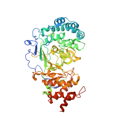
5A7R - PubMed Abstract:
Poly(ADP-ribosyl)ation is a common post-translational modification that mediates a wide variety of cellular processes including DNA damage repair, chromatin regulation, transcription, and apoptosis. The difficulty associated with accessing poly(ADP-ribose) (PAR) in a homogeneous form has been an impediment to understanding the interactions of PAR with poly(ADP-ribose) glycohydrolase (PARG) and other binding proteins. Here we describe the chemical synthesis of the ADP-ribose dimer, and we use this compound to obtain the first human PARG substrate-enzyme cocrystal structure. Chemical synthesis of PAR is an attractive alternative to traditional enzymatic synthesis and fractionation, allowing access to products such as dimeric ADP-ribose, which has been detected but never isolated from natural sources. Additionally, we describe the synthesis of an alkynylated dimer and demonstrate that this compound can be used to synthesize PAR probes including biotin and fluorophore-labeled compounds. The fluorescently labeled ADP-ribose dimer was then utilized in a general fluorescence polarization-based PAR-protein binding assay. Finally, we use intermediates of our synthesis to access various PAR fragments, and evaluation of these compounds as substrates for PARG reveals the minimal features for substrate recognition and enzymatic cleavage. Homogeneous PAR oligomers and unnatural variants produced from chemical synthesis will allow for further detailed structural and biochemical studies on the interaction of PAR with its many protein binding partners.
- †Department of Chemistry, Roger Adams Laboratory, University of Illinois, 600 South Mathews, Urbana, Illinois 61801, United States.
Organizational Affiliation: