5AUN | pdb_00005aun

Crystal structure of the HypAB-Ni complex


Experimental Data Snapshot

  • Method: X-RAY DIFFRACTION
  • Resolution: 1.63 Å
  • R-Value Free: 
    0.213 (Depositor), 0.216 (DCC) 
  • R-Value Work: 
    0.180 (Depositor), 0.183 (DCC) 
  • R-Value Observed: 
    0.182 (Depositor) 

Starting Models: experimental
View more details

wwPDB Validation 3D Report Full Report

Validation slider image for 5AUN

Ligand Structure Quality Assessment 


This is version 1.3 of the entry. See complete history

Literature

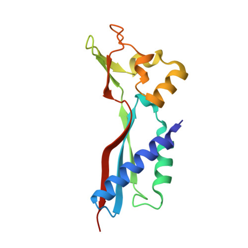
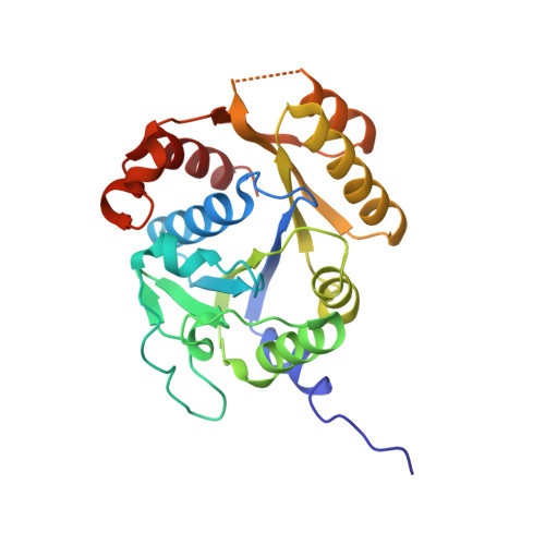
Structural basis of a Ni acquisition cycle for [NiFe] hydrogenase by Ni-metallochaperone HypA and its enhancer

Watanabe, S.Kawashima, T.Nishitani, Y.Kanai, T.Wada, T.Inaba, K.Atomi, H.Imanaka, T.Miki, K.

(2015) Proc Natl Acad Sci U S A 112: 7701-7706

  • DOI: https://doi.org/10.1073/pnas.1503102112
  • Primary Citation Related Structures: 
    5AUN, 5AUO, 5AUP, 5AUQ

  • PubMed Abstract: 

    The Ni atom at the catalytic center of [NiFe] hydrogenases is incorporated by a Ni-metallochaperone, HypA, and a GTPase/ATPase, HypB. We report the crystal structures of the transient complex formed between HypA and ATPase-type HypB (HypBAT) with Ni ions. Transient association between HypA and HypBAT is controlled by the ATP hydrolysis cycle of HypBAT, which is accelerated by HypA. Only the ATP-bound form of HypBAT can interact with HypA and induces drastic conformational changes of HypA. Consequently, upon complex formation, a conserved His residue of HypA comes close to the N-terminal conserved motif of HypA and forms a Ni-binding site, to which a Ni ion is bound with a nearly square-planar geometry. The Ni binding site in the HypABAT complex has a nanomolar affinity (Kd = 7 nM), which is in contrast to the micromolar affinity (Kd = 4 µM) observed with the isolated HypA. The ATP hydrolysis and Ni binding cause conformational changes of HypBAT, affecting its association with HypA. These findings indicate that HypA and HypBAT constitute an ATP-dependent Ni acquisition cycle for [NiFe]-hydrogenase maturation, wherein HypBAT functions as a metallochaperone enhancer and considerably increases the Ni-binding affinity of HypA.


  • Organizational Affiliation
    • Department of Chemistry, Graduate School of Science, Kyoto University, Sakyo-ku, Kyoto 606-8502, Japan; Institute of Multidisciplinary Research for Advanced Materials, Tohoku University, Katahira 2-1-1, Aoba-ku, Sendai 980-8577, Japan;

Macromolecule Content 

  • Total Structure Weight: 44.33 kDa 
  • Atom Count: 3,384 
  • Modeled Residue Count: 376 
  • Deposited Residue Count: 387 
  • Unique protein chains: 2

Macromolecules

Find similar proteins by:|  3D Structure
Entity ID: 1
MoleculeChains  Sequence LengthOrganismDetailsImage
Probable hydrogenase nickel incorporation protein HypA139Thermococcus kodakarensis KOD1Mutation(s): 0 
Gene Names: hypATK2008
UniProt
Find proteins for Q5JIH3 (Thermococcus kodakarensis (strain ATCC BAA-918 / JCM 12380 / KOD1))
Explore Q5JIH3 
Go to UniProtKB:  Q5JIH3
Entity Groups
Sequence Clusters30% Identity50% Identity70% Identity90% Identity95% Identity100% Identity
UniProt GroupQ5JIH3
Sequence Annotations
Expand
Reference Sequence
Find similar proteins by:|  3D Structure
Entity ID: 2
MoleculeChains  Sequence LengthOrganismDetailsImage
ATPase involved in chromosome partitioning, ParA/MinD family, Mrp homolog248Thermococcus kodakarensis KOD1Mutation(s): 0 
Gene Names: TK2007
UniProt
Find proteins for Q5JIH4 (Thermococcus kodakarensis (strain ATCC BAA-918 / JCM 12380 / KOD1))
Explore Q5JIH4 
Go to UniProtKB:  Q5JIH4
Entity Groups
Sequence Clusters30% Identity50% Identity70% Identity90% Identity95% Identity100% Identity
UniProt GroupQ5JIH4
Sequence Annotations
Expand
Reference Sequence

Small Molecules

Ligands 7 Unique
IDChains Name / Formula / InChI Key2D Diagram3D Interactions
ADP

Query on ADP


Download:Ideal Coordinates CCD File
J [auth B]ADENOSINE-5'-DIPHOSPHATE
C10 H15 N5 O10 P2
XTWYTFMLZFPYCI-KQYNXXCUSA-N
GOL

Query on GOL


Download:Ideal Coordinates CCD File
I [auth A],
M [auth B]
GLYCEROL
C3 H8 O3
PEDCQBHIVMGVHV-UHFFFAOYSA-N
ZN

Query on ZN


Download:Ideal Coordinates CCD File
C [auth A]ZINC ION
Zn
PTFCDOFLOPIGGS-UHFFFAOYSA-N
URE

Query on URE


Download:Ideal Coordinates CCD File
E [auth A],
F [auth A],
G [auth A]
UREA
C H4 N2 O
XSQUKJJJFZCRTK-UHFFFAOYSA-N
NI

Query on NI


Download:Ideal Coordinates CCD File
D [auth A]NICKEL (II) ION
Ni
VEQPNABPJHWNSG-UHFFFAOYSA-N
CL

Query on CL


Download:Ideal Coordinates CCD File
H [auth A]CHLORIDE ION
Cl
VEXZGXHMUGYJMC-UHFFFAOYSA-M
MG

Query on MG


Download:Ideal Coordinates CCD File
K [auth B],
L [auth B]
MAGNESIUM ION
Mg
JLVVSXFLKOJNIY-UHFFFAOYSA-N

Experimental Data & Validation

Experimental Data

  • Method: X-RAY DIFFRACTION
  • Resolution: 1.63 Å
  • R-Value Free:  0.213 (Depositor), 0.216 (DCC) 
  • R-Value Work:  0.180 (Depositor), 0.183 (DCC) 
  • R-Value Observed: 0.182 (Depositor) 
Space Group: P 21 21 2
Unit Cell:
Length ( Å )Angle ( ˚ )
a = 74.72α = 90
b = 82.744β = 90
c = 65.128γ = 90
Software Package:
Software NamePurpose
PHENIXrefinement
HKL-2000data reduction
HKL-2000data scaling
MOLREPphasing

Structure Validation

View Full Validation Report


Ligand Structure Quality Assessment 


Entry History 

Deposition Data

Revision History  (Full details and data files)

  • Version 1.0: 2015-06-24
    Type: Initial release
  • Version 1.1: 2015-07-08
    Changes: Database references
  • Version 1.2: 2020-02-19
    Changes: Advisory, Data collection, Database references, Derived calculations, Source and taxonomy, Structure summary
  • Version 1.3: 2023-11-08
    Changes: Data collection, Database references, Derived calculations, Refinement description