


Nucleotide binding to variants of the HSP70-NBD.
Narayanan, D., Pflug, A., Christopeit, T., Kyomuhendo, P., Engh, R.A.To be published.
Experimental Data Snapshot
Starting Model: experimental
View more details
Entity ID: 1 | |||||
|---|---|---|---|---|---|
| Molecule | Chains | Sequence Length | Organism | Details | Image |
| Heat shock 70 kDa protein 1A | 388 | Homo sapiens | Mutation(s): 1 Gene Names: HSPA1A, HSPA1, HSX70 | 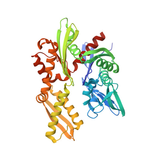 | |
UniProt & NIH Common Fund Data Resources | |||||
PHAROS: P0DMV8 | |||||
Entity Groups | |||||
| Sequence Clusters | 30% Identity50% Identity70% Identity90% Identity95% Identity100% Identity | ||||
| UniProt Group | P0DMV8 | ||||
Sequence AnnotationsExpand | |||||
Reference Sequence | |||||
| Ligands 6 Unique | |||||
|---|---|---|---|---|---|
| ID | Chains | Name / Formula / InChI Key | 2D Diagram | 3D Interactions | |
| ADP Download:Ideal Coordinates CCD File | B [auth A] | ADENOSINE-5'-DIPHOSPHATE C10 H15 N5 O10 P2 XTWYTFMLZFPYCI-KQYNXXCUSA-N |  | ||
| EPE Download:Ideal Coordinates CCD File | I [auth A] | 4-(2-HYDROXYETHYL)-1-PIPERAZINE ETHANESULFONIC ACID C8 H18 N2 O4 S JKMHFZQWWAIEOD-UHFFFAOYSA-N |  | ||
| PO4 Download:Ideal Coordinates CCD File | C [auth A], D [auth A], E [auth A] | PHOSPHATE ION O4 P NBIIXXVUZAFLBC-UHFFFAOYSA-K |  | ||
| CL Download:Ideal Coordinates CCD File | J [auth A] | CHLORIDE ION Cl VEXZGXHMUGYJMC-UHFFFAOYSA-M |  | ||
| MG Download:Ideal Coordinates CCD File | F [auth A] | MAGNESIUM ION Mg JLVVSXFLKOJNIY-UHFFFAOYSA-N |  | ||
| NA Download:Ideal Coordinates CCD File | G [auth A], H [auth A] | SODIUM ION Na FKNQFGJONOIPTF-UHFFFAOYSA-N |  | ||
| Length ( Å ) | Angle ( ˚ ) |
|---|---|
| a = 46.037 | α = 90 |
| b = 63.327 | β = 90 |
| c = 143.798 | γ = 90 |
| Software Name | Purpose |
|---|---|
| PHENIX | refinement |
| Coot | model building |
| MOLREP | phasing |
| Aimless | data scaling |
| XDS | data reduction |
| Funding Organization | Location | Grant Number |
|---|---|---|
| FRINAT | Norway | 191303 |


