5CA2 | pdb_00005ca2

CONFORMATIONAL MOBILITY OF HIS-64 IN THE THR-200 (RIGHT ARROW) SER MUTANT OF HUMAN CARBONIC ANHYDRASE II


Experimental Data Snapshot

  • Method: X-RAY DIFFRACTION
  • Resolution: 2.10 Å
  • R-Value Observed: 
    0.160 (Depositor) 

wwPDB Validation 3D Report Full Report

Validation slider image for 5CA2

This is version 1.4 of the entry. See complete history

Literature

Conformational mobility of His-64 in the Thr-200----Ser mutant of human carbonic anhydrase II.

Krebs, J.F.Fierke, C.A.Alexander, R.S.Christianson, D.W.

(1991) Biochemistry 30: 9153-9160

  • DOI: https://doi.org/10.1021/bi00102a005
  • Primary Citation Related Structures: 
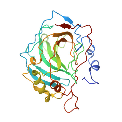
    5CA2

  • PubMed Abstract: 

    The three-dimensional structure of the Thr-200----Ser (T200S) mutant of human carbonic anhydrase II (CAII) has been determined by X-ray crystallographic methods at 2.1-A resolution. This particular mutant of CAII exhibits CO2 hydrase activity that is comparable to that of the wild-type enzyme with a 2-fold stabilization of the E.HCO3- complex and esterase activity that is 4-fold greater than that of the wild-type enzyme. The structure of the mutant enzyme reveals no significant local changes accompanying the conservative T200S substitution, but an important nonlocal structural change is evident: the side chain of catalytic residue His-64 rotates away from the active site by 105 degrees about chi 1 and apparently displaces a water molecule. The displaced water molecule is present in the wild-type enzyme; however, the electron density into which this water is built is interpretable as an alternate conformation of His-64 with 10-20% occupancy. The rate constants for proton transfer from the zinc-water ligand to His-64 and from His-64 to bulk solvent are maintained in the T200S variant; therefore, if His-64 is conformationally mobile about chi 1 and/or chi 2 during catalysis, compensatory changes in solvent configuration must sustain efficient proton transfer.


  • Organizational Affiliation
    • Department of Biochemistry, Duke University Medical Center, Durham, North Carolina 27710.

Macromolecule Content 

  • Total Structure Weight: 29.54 kDa 
  • Atom Count: 2,149 
  • Modeled Residue Count: 255 
  • Deposited Residue Count: 260 
  • Unique protein chains: 1

Macromolecules

Find similar proteins by:|  3D Structure
Entity ID: 1
MoleculeChains  Sequence LengthOrganismDetailsImage
CARBONIC ANHYDRASE II260Homo sapiensMutation(s): 0 
EC: 4.2.1.1 (PDB Primary Data), 4.2.1.69 (UniProt)
UniProt & NIH Common Fund Data Resources
Find proteins for P00918 (Homo sapiens)
Explore P00918 
Go to UniProtKB:  P00918
PHAROS:  P00918
GTEx:  ENSG00000104267 
Entity Groups
Sequence Clusters30% Identity50% Identity70% Identity90% Identity95% Identity100% Identity
UniProt GroupP00918
Sequence Annotations
Expand
Reference Sequence

Small Molecules

Ligands 2 Unique
IDChains Name / Formula / InChI Key2D Diagram3D Interactions
HG

Query on HG


Download:Ideal Coordinates CCD File
C [auth A]MERCURY (II) ION
Hg
BQPIGGFYSBELGY-UHFFFAOYSA-N
ZN

Query on ZN


Download:Ideal Coordinates CCD File
B [auth A]ZINC ION
Zn
PTFCDOFLOPIGGS-UHFFFAOYSA-N

Experimental Data & Validation

Experimental Data

  • Method: X-RAY DIFFRACTION
  • Resolution: 2.10 Å
  • R-Value Observed: 0.160 (Depositor) 
Space Group: P 1 21 1
Unit Cell:
Length ( Å )Angle ( ˚ )
a = 42.7α = 90
b = 41.7β = 104.6
c = 73γ = 90
Software Package:
Software NamePurpose
PROLSQrefinement

Structure Validation

View Full Validation Report


Entry History 

Deposition Data

Revision History  (Full details and data files)

  • Version 1.0: 1992-10-31
    Type: Initial release
  • Version 1.1: 2008-03-25
    Changes: Version format compliance
  • Version 1.2: 2011-07-13
    Changes: Version format compliance
  • Version 1.3: 2017-11-29
    Changes: Derived calculations, Other
  • Version 1.4: 2024-03-06
    Changes: Data collection, Database references, Derived calculations