5CI8 | pdb_00005ci8

Crystal structure of human Tob in complex with inhibitor fragment 1


Experimental Data Snapshot

  • Method: X-RAY DIFFRACTION
  • Resolution: 2.33 Å
  • R-Value Free: 
    0.285 (Depositor), 0.298 (DCC) 
  • R-Value Work: 
    0.232 (Depositor), 0.244 (DCC) 
  • R-Value Observed: 
    0.238 (Depositor) 

wwPDB Validation 3D Report Full Report

Validation slider image for 5CI8

Ligand Structure Quality Assessment 


This is version 1.3 of the entry. See complete history

Literature

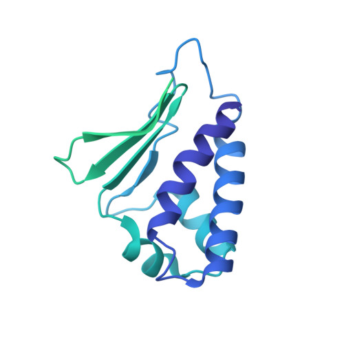
Structural basis for inhibition of the Tob-CNOT7 interaction by a fragment screening approach

Bai, Y.Tashiro, S.Nagatoishi, S.Suzuki, T.Yan, D.Liu, R.Tsumoto, K.Bartlam, M.Yamamoto, T.

(2015) Protein Cell 6: 924-928

  • DOI: https://doi.org/10.1007/s13238-015-0225-6
  • Primary Citation Related Structures: 
    5CI8, 5CI9

  • Organizational Affiliation
    • State Key Laboratory of Medicinal Chemical Biology, Nankai University, Tianjin, 300071, China.

Macromolecule Content 

  • Total Structure Weight: 38.65 kDa 
  • Atom Count: 981 
  • Modeled Residue Count: 117 
  • Deposited Residue Count: 347 
  • Unique protein chains: 1

Macromolecules

Find similar proteins by:|  3D Structure
Entity ID: 1
MoleculeChains  Sequence LengthOrganismDetailsImage
Protein Tob1347Homo sapiensMutation(s): 0 
Gene Names: TOB1TOBTROB1
UniProt & NIH Common Fund Data Resources
Find proteins for P50616 (Homo sapiens)
Explore P50616 
Go to UniProtKB:  P50616
PHAROS:  P50616
GTEx:  ENSG00000141232 
Entity Groups
Sequence Clusters30% Identity50% Identity70% Identity90% Identity95% Identity100% Identity
UniProt GroupP50616
Sequence Annotations
Expand
Reference Sequence

Small Molecules

Ligands 1 Unique
IDChains Name / Formula / InChI Key2D Diagram3D Interactions
53Y

Query on 53Y


Download:Ideal Coordinates CCD File
B [auth A]pyrrolo[1,2-a]quinoxalin-4(5H)-one
C11 H8 N2 O
LINHQLFBBDHSEJ-UHFFFAOYSA-N

Experimental Data & Validation

Experimental Data

  • Method: X-RAY DIFFRACTION
  • Resolution: 2.33 Å
  • R-Value Free:  0.285 (Depositor), 0.298 (DCC) 
  • R-Value Work:  0.232 (Depositor), 0.244 (DCC) 
  • R-Value Observed: 0.238 (Depositor) 
Space Group: P 61 2 2
Unit Cell:
Length ( Å )Angle ( ˚ )
a = 64.975α = 90
b = 64.975β = 90
c = 163.677γ = 120
Software Package:
Software NamePurpose
PHENIXrefinement
HKL-2000data reduction
HKL-2000data scaling
PHASERphasing

Structure Validation

View Full Validation Report


Ligand Structure Quality Assessment 


Entry History 

& Funding Information

Deposition Data


Funding OrganizationLocationGrant Number
Ministry of Science and TechnologyChina2014CB560709
Ministry of Education, Culture, Sports, Science and Technology (Japan)Japan--
KAKENHIJapan25121734

Revision History  (Full details and data files)

  • Version 1.0: 2015-11-18
    Type: Initial release
  • Version 1.1: 2015-12-09
    Changes: Database references
  • Version 1.2: 2025-04-09
    Changes: Author supporting evidence, Data collection, Database references, Derived calculations, Structure summary
  • Version 1.3: 2025-09-17
    Changes: Advisory, Derived calculations