5CUS

Crystal Structure of sErbB3-Fab3379 Complex


Experimental Data Snapshot

  • Method: X-RAY DIFFRACTION
  • Resolution: 3.20 Å
  • R-Value Free: 0.278 
  • R-Value Work: 0.252 
  • R-Value Observed: 0.254 

wwPDB Validation   3D Report Full Report


Ligand Structure Quality Assessment 


This is version 1.5 of the entry. See complete history


Literature

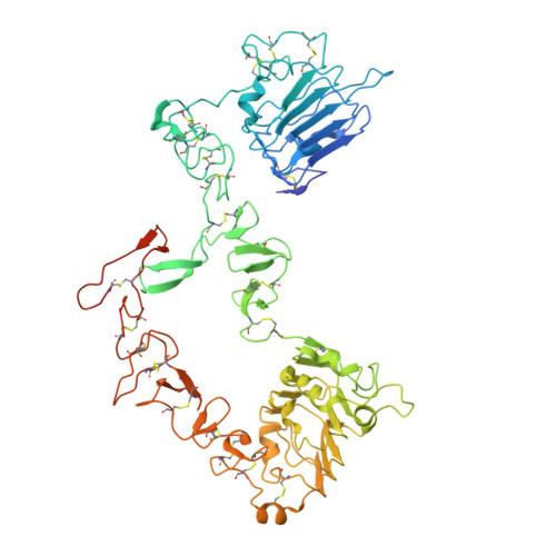
Inhibition of ErbB3 by a monoclonal antibody that locks the extracellular domain in an inactive configuration.

Lee, S.Greenlee, E.B.Amick, J.R.Ligon, G.F.Lillquist, J.S.Natoli, E.J.Hadari, Y.Alvarado, D.Schlessinger, J.

(2015) Proc Natl Acad Sci U S A 112: 13225-13230

  • DOI: https://doi.org/10.1073/pnas.1518361112
  • Primary Citation of Related Structures:  
    5CUS

  • PubMed Abstract: 

    ErbB3 (HER3) is a member of the EGF receptor (EGFR) family of receptor tyrosine kinases, which, unlike the other three family members, contains a pseudo kinase in place of a tyrosine kinase domain. In cancer, ErbB3 activation is driven by a ligand-dependent mechanism through the formation of heterodimers with EGFR, ErbB2, or ErbB4 or via a ligand-independent process through heterodimerization with ErbB2 overexpressed in breast tumors or other cancers. Here we describe the crystal structure of the Fab fragment of an antagonistic monoclonal antibody KTN3379, currently in clinical development in human cancer patients, in complex with the ErbB3 extracellular domain. The structure reveals a unique allosteric mechanism for inhibition of ligand-dependent or ligand-independent ErbB3-driven cancers by binding to an epitope that locks ErbB3 in an inactive conformation. Given the similarities in the mechanism of ErbB receptor family activation, these findings could facilitate structure-based design of antibodies that inhibit EGFR and ErbB4 by an allosteric mechanism.


  • Organizational Affiliation

    Department of Pharmacology, Yale School of Medicine, New Haven, CT 06520;


Macromolecules
Find similar proteins by:  (by identity cutoff)  |  3D Structure
Entity ID: 1
MoleculeChains Sequence LengthOrganismDetailsImage
Receptor tyrosine-protein kinase erbB-3
A, B, C, D
627Homo sapiensMutation(s): 0 
Gene Names: ERBB3HER3
EC: 2.7.10.1
UniProt & NIH Common Fund Data Resources
Find proteins for P21860 (Homo sapiens)
Explore P21860 
Go to UniProtKB:  P21860
PHAROS:  P21860
GTEx:  ENSG00000065361 
Entity Groups  
Sequence Clusters30% Identity50% Identity70% Identity90% Identity95% Identity100% Identity
UniProt GroupP21860
Sequence Annotations
Expand
  • Reference Sequence
Find similar proteins by:  (by identity cutoff)  |  3D Structure
Entity ID: 2
MoleculeChains Sequence LengthOrganismDetailsImage
IgG H chainE [auth H],
G [auth I],
H [auth J],
I [auth K]
221Homo sapiensMutation(s): 0 
UniProt
Find proteins for S6B291 (Homo sapiens)
Explore S6B291 
Go to UniProtKB:  S6B291
Entity Groups  
Sequence Clusters30% Identity50% Identity70% Identity90% Identity95% Identity100% Identity
UniProt GroupS6B291
Sequence Annotations
Expand
  • Reference Sequence
Find similar proteins by:  (by identity cutoff)  |  3D Structure
Entity ID: 3
MoleculeChains Sequence LengthOrganismDetailsImage
Fab LC region of KTN3379F [auth L],
J [auth M],
K [auth N],
L [auth O]
216Homo sapiensMutation(s): 0 
Entity Groups  
Sequence Clusters30% Identity50% Identity70% Identity90% Identity95% Identity100% Identity
Sequence Annotations
Expand
  • Reference Sequence
Small Molecules
Ligands 1 Unique
IDChains Name / Formula / InChI Key2D Diagram3D Interactions
NAG
Query on NAG

Download Ideal Coordinates CCD File 
M [auth A]
N [auth A]
O [auth B]
P [auth B]
Q [auth C]
M [auth A],
N [auth A],
O [auth B],
P [auth B],
Q [auth C],
R [auth C],
S [auth C],
T [auth D],
U [auth D],
V [auth D],
W [auth M]
2-acetamido-2-deoxy-beta-D-glucopyranose
C8 H15 N O6
OVRNDRQMDRJTHS-FMDGEEDCSA-N
Experimental Data & Validation

Experimental Data

  • Method: X-RAY DIFFRACTION
  • Resolution: 3.20 Å
  • R-Value Free: 0.278 
  • R-Value Work: 0.252 
  • R-Value Observed: 0.254 
  • Space Group: P 1
Unit Cell:
Length ( Å )Angle ( ˚ )
a = 84.378α = 87.1
b = 127.141β = 85.54
c = 138.078γ = 89.92
Software Package:
Software NamePurpose
PHENIXrefinement
XDSdata scaling
PHASERphasing
XDSdata reduction

Structure Validation

View Full Validation Report


Ligand Structure Quality Assessment 


Entry History 

Deposition Data

Revision History  (Full details and data files)

  • Version 1.0: 2015-10-14
    Type: Initial release
  • Version 1.1: 2015-10-28
    Changes: Database references
  • Version 1.2: 2015-11-04
    Changes: Database references
  • Version 1.3: 2017-11-22
    Changes: Advisory, Database references, Derived calculations, Refinement description
  • Version 1.4: 2020-07-29
    Type: Remediation
    Reason: Carbohydrate remediation
    Changes: Data collection, Derived calculations, Structure summary
  • Version 1.5: 2023-09-27
    Changes: Advisory, Data collection, Database references, Refinement description, Structure summary