


Neisseria meningitidis 3 deoxy-D-arabino-heptulosonate 7-phosphate synthase Glu176Gln variant at 1.87 Angstroms resolution
Heyes, L.C.To be published.
Experimental Data Snapshot
Entity ID: 1 | |||||
|---|---|---|---|---|---|
| Molecule | Chains | Sequence Length | Organism | Details | Image |
| Phospho-2-dehydro-3-deoxyheptonate aldolase | 351 | Neisseria meningitidis MC58 | Mutation(s): 1 Gene Names: aroG, NMB0307 EC: 2.5.1.54 | 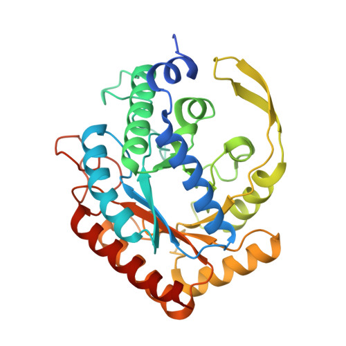 | |
UniProt | |||||
Entity Groups | |||||
| Sequence Clusters | 30% Identity50% Identity70% Identity90% Identity95% Identity100% Identity | ||||
| UniProt Group | Q9K169 | ||||
Sequence AnnotationsExpand | |||||
Reference Sequence | |||||
| Ligands 4 Unique | |||||
|---|---|---|---|---|---|
| ID | Chains | Name / Formula / InChI Key | 2D Diagram | 3D Interactions | |
| PEP Download:Ideal Coordinates CCD File | F [auth A], I [auth B], M [auth C], Q [auth D] | PHOSPHOENOLPYRUVATE C3 H5 O6 P DTBNBXWJWCWCIK-UHFFFAOYSA-N |  | ||
| SO4 Download:Ideal Coordinates CCD File | G [auth A], S [auth D] | SULFATE ION O4 S QAOWNCQODCNURD-UHFFFAOYSA-L |  | ||
| EDO Download:Ideal Coordinates CCD File | J [auth B], K [auth B], N [auth C], O [auth C], R [auth D] | 1,2-ETHANEDIOL C2 H6 O2 LYCAIKOWRPUZTN-UHFFFAOYSA-N |  | ||
| MN Download:Ideal Coordinates CCD File | E [auth A], H [auth B], L [auth C], P [auth D] | MANGANESE (II) ION Mn WAEMQWOKJMHJLA-UHFFFAOYSA-N |  | ||
| Length ( Å ) | Angle ( ˚ ) |
|---|---|
| a = 73.54 | α = 90 |
| b = 135.546 | β = 96.27 |
| c = 76.009 | γ = 90 |
| Software Name | Purpose |
|---|---|
| REFMAC | refinement |
| XDS | data reduction |
| Aimless | data scaling |
| PHASER | phasing |


