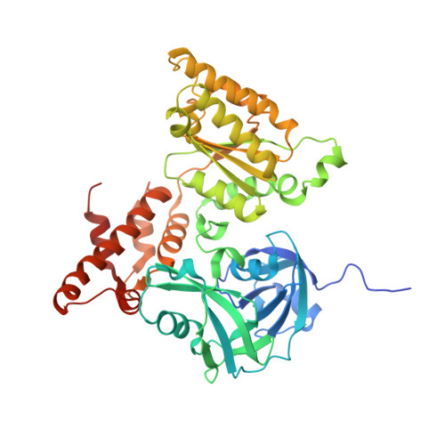
5DYG

Structure of p97 N-D1 L198W mutant in complex with ADP


Experimental Data Snapshot

  • Method: X-RAY DIFFRACTION
  • Resolution: 2.20 Å
  • R-Value Free: 0.258 
  • R-Value Work: 0.200 
  • R-Value Observed: 0.203 

wwPDB Validation   3D Report Full Report


Ligand Structure Quality Assessment 


This is version 1.1 of the entry. See complete history


Literature

Role of the D1-D2 Linker of Human VCP/p97 in the Asymmetry and ATPase Activity of the D1-domain.

Tang, W.K.Xia, D.

(2016) Sci Rep 6: 20037-20037

  • DOI: https://doi.org/10.1038/srep20037
  • Primary Citation of Related Structures:  
    5DYG, 5DYI

  • PubMed Abstract: 

    Human AAA(+) protein p97 consists of an N-domain and two tandem ATPase domains D1 and D2, which are connected by the N-D1 and the D1-D2 linkers. Inclusion of the D1-D2 linker, a 22-amino acid peptide, at the end of p97 N-D1 truncate has been shown to activate ATP hydrolysis of its D1-domain, although the mechanism of activation remains unclear. Here, we identify the N-terminal half of this linker, highly conserved from human to fungi, is essential for the ATPase activation. By analyzing available crystal structures, we observed that the D1-D2 linker is capable of inducing asymmetry in subunit association into a p97 hexamer. This observation is reinforced by two new crystal structures, determined in the present work. The effect of D1-D2 linker on the ATPase activity of the D1-domain is correlated to the side-chain conformation of residue R359, a trans-acting arginine-finger residue essential for ATP hydrolysis of the D1-domain. The activation in D1-domain ATPase activity by breaking perfect six-fold symmetry implies functional importance of asymmetric association of p97 subunits, the extent of which can be determined quantitatively by the metric Asymmetric Index.


  • Organizational Affiliation

    Laboratory of Cell Biology, Center for Cancer Research, National Cancer Institute, National Institutes of Health, Bethesda, MD 20892, USA.


Macromolecules
Find similar proteins by:  (by identity cutoff)  |  3D Structure
Entity ID: 1
MoleculeChains Sequence LengthOrganismDetailsImage
Transitional endoplasmic reticulum ATPase468Homo sapiensMutation(s): 1 
Gene Names: VCP
EC: 3.6.4.6
UniProt & NIH Common Fund Data Resources
Find proteins for P55072 (Homo sapiens)
Explore P55072 
Go to UniProtKB:  P55072
PHAROS:  P55072
GTEx:  ENSG00000165280 
Entity Groups  
Sequence Clusters30% Identity50% Identity70% Identity90% Identity95% Identity100% Identity
UniProt GroupP55072
Sequence Annotations
Expand
  • Reference Sequence
Small Molecules
Ligands 1 Unique
IDChains Name / Formula / InChI Key2D Diagram3D Interactions
ADP
Query on ADP

Download Ideal Coordinates CCD File 
B [auth A]ADENOSINE-5'-DIPHOSPHATE
C10 H15 N5 O10 P2
XTWYTFMLZFPYCI-KQYNXXCUSA-N
Experimental Data & Validation

Experimental Data

  • Method: X-RAY DIFFRACTION
  • Resolution: 2.20 Å
  • R-Value Free: 0.258 
  • R-Value Work: 0.200 
  • R-Value Observed: 0.203 
  • Space Group: P 6 2 2
Unit Cell:
Length ( Å )Angle ( ˚ )
a = 145.584α = 90
b = 145.584β = 90
c = 84.002γ = 120
Software Package:
Software NamePurpose
REFMACrefinement
DENZOdata reduction
SCALEPACKdata scaling
MOLREPphasing

Structure Validation

View Full Validation Report


Ligand Structure Quality Assessment 


Entry History 

Deposition Data

Revision History  (Full details and data files)

  • Version 1.0: 2016-02-10
    Type: Initial release
  • Version 1.1: 2024-03-06
    Changes: Data collection, Database references, Derived calculations