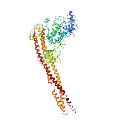
5EBZ

Crystal structure of human IKK1


Experimental Data Snapshot

  • Method: X-RAY DIFFRACTION
  • Resolution: 4.50 Å
  • R-Value Free: 0.276 
  • R-Value Work: 0.238 
  • R-Value Observed: 0.238 

wwPDB Validation   3D Report Full Report


This is version 2.1 of the entry. See complete history


Literature

Structural Basis for the Activation of IKK1/ alpha.

Polley, S.Passos, D.O.Huang, D.B.Mulero, M.C.Mazumder, A.Biswas, T.Verma, I.M.Lyumkis, D.Ghosh, G.

(2016) Cell Rep 17: 1907-1914

  • DOI: https://doi.org/10.1016/j.celrep.2016.10.067
  • Primary Citation of Related Structures:  
    5EBZ, 5TQW, 5TQX, 5TQY

  • PubMed Abstract: 

    Distinct signaling pathways activate the NF-κB family of transcription factors. The canonical NF-κB-signaling pathway is mediated by IκB kinase 2/β (IKK2/β), while the non-canonical pathway depends on IKK1/α. The structural and biochemical bases for distinct signaling by these otherwise highly similar IKKs are unclear. We report single-particle cryoelectron microscopy (cryo-EM) and X-ray crystal structures of human IKK1 in dimeric (∼150 kDa) and hexameric (∼450 kDa) forms. The hexamer, which is the representative form in the crystal but comprises only ∼2% of the particles in solution by cryo-EM, is a trimer of IKK1 dimers. While IKK1 hexamers are not detectable in cells, the surface that supports hexamer formation is critical for IKK1-dependent cellular processing of p100 to p52, the hallmark of non-canonical NF-κB signaling. Comparison of this surface to that in IKK2 indicates significant divergence, and it suggests a fundamental role for this surface in signaling by these kinases through distinct pathways.


  • Organizational Affiliation

    Department of Chemistry & Biochemistry, University of California San Diego, La Jolla, CA 92093, USA; Laboratory of Genetics and Helmsley Center for Genomic Medicine, The Salk Institute for Biological Studies, La Jolla, CA 92037, USA.


Macromolecules
Find similar proteins by:  (by identity cutoff)  |  3D Structure
Entity ID: 1
MoleculeChains Sequence LengthOrganismDetailsImage
Inhibitor of nuclear factor kappa-B kinase subunit alpha
A, B, C, D, E
655Homo sapiensMutation(s): 3 
Gene Names: CHUKIKKATCF16
EC: 2.7.11.10
UniProt & NIH Common Fund Data Resources
Find proteins for O15111 (Homo sapiens)
Explore O15111 
Go to UniProtKB:  O15111
PHAROS:  O15111
GTEx:  ENSG00000213341 
Entity Groups  
Sequence Clusters30% Identity50% Identity70% Identity90% Identity95% Identity100% Identity
UniProt GroupO15111
Sequence Annotations
Expand
  • Reference Sequence
Oligosaccharides

Help

Entity ID: 3
MoleculeChains Length2D Diagram Glycosylation3D Interactions
[(2S,3R,4S,5S,6R)-6-(hydroxymethyl)-2,4-bis(oxidanyl)-5-oxidanylsulfanyloxy-oxan-3-yl] hydrogen sulfate-(1-6)-(2S,3R,4R,5S,6R)-6-(hydroxymethyl)-5-oxidanylsulfanyloxy-oxane-2,3,4-triol-(1-6)-2-O-sulfo-alpha-D-glucopyranose-(1-6)-[(2R,3R,4R,5R,6S)-2-(hydroxymethyl)-5,6-bis(oxidanyl)-3-oxidanylsulfanyloxy-oxan-4-yl] hydrogen sulfate-(1-6)-[(2S,3R,4S,5R,6R)-6-(hydroxymethyl)-2,5-bis(oxidanyl)-4-oxidanylsulfanyloxy-oxan-3-yl] hydrogen sulfate-(1-6)-[(2R,3R,4R,5R,6S)-2-(hydroxymethyl)-5,6-bis(oxidanyl)-3-oxidanylsulfanyloxy-oxan-4-yl] hydrogen sulfate-(1-6)-2,4-di-O-sulfo-alpha-D-glucopyranoseCA [auth c],
IA [auth i],
O,
T
7N/AN/A
Glycosylation Resources
GlyTouCan:  G51196UN
GlyCosmos:  G51196UN
Small Molecules
Ligands 1 Unique
IDChains Name / Formula / InChI Key2D Diagram3D Interactions
5TL
Query on 5TL

Download Ideal Coordinates CCD File 
OA [auth A]
PA [auth B]
QA [auth C]
RA [auth D]
SA [auth E]
2-azanyl-5-phenyl-3-(4-sulfamoylphenyl)benzamide
C19 H17 N3 O3 S
IVJPEEHVYKDQEO-UHFFFAOYSA-N
Binding Affinity Annotations 
IDSourceBinding Affinity
5TL BindingDB:  5EBZ IC50: 1000 (nM) from 1 assay(s)
Experimental Data & Validation

Experimental Data

  • Method: X-RAY DIFFRACTION
  • Resolution: 4.50 Å
  • R-Value Free: 0.276 
  • R-Value Work: 0.238 
  • R-Value Observed: 0.238 
  • Space Group: P 1 21 1
Unit Cell:
Length ( Å )Angle ( ˚ )
a = 174.51α = 90
b = 186.94β = 98.84
c = 275.83γ = 90
Software Package:
Software NamePurpose
CNSrefinement
HKL-2000data collection
HKL-2000data scaling
PDB_EXTRACTdata extraction
HKL-2000data reduction
MOLREPphasing

Structure Validation

View Full Validation Report


Entry History & Funding Information

Deposition Data


Funding OrganizationLocationGrant Number
National Institutes of Health/National Cancer Institute (NIH/NCI)United StatesCA141722

Revision History  (Full details and data files)

  • Version 1.0: 2016-11-02
    Type: Initial release
  • Version 1.1: 2016-12-21
    Changes: Database references
  • Version 1.2: 2017-09-06
    Changes: Author supporting evidence
  • Version 1.3: 2019-12-04
    Changes: Author supporting evidence, Data collection
  • Version 2.0: 2020-07-29
    Type: Remediation
    Reason: Carbohydrate remediation
    Changes: Atomic model, Data collection, Derived calculations, Structure summary
  • Version 2.1: 2024-03-06
    Changes: Data collection, Database references, Derived calculations, Structure summary