5EJM | pdb_00005ejm

ThDP-Mn2+ complex of R413A variant of EcMenD soaked with 2-ketoglutarate for 35 min


Experimental Data Snapshot

  • Method: X-RAY DIFFRACTION
  • Resolution: 1.72 Å
  • R-Value Free: 
    0.192 (Depositor), 0.192 (DCC) 
  • R-Value Work: 
    0.164 (Depositor), 0.165 (DCC) 
  • R-Value Observed: 
    0.165 (Depositor) 

wwPDB Validation 3D Report Full Report

Validation slider image for 5EJM

Ligand Structure Quality Assessment 


This is version 1.2 of the entry. See complete history

Literature

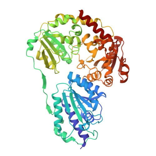
Two active site arginines are critical determinants of substrate binding and catalysis in MenD, a thiamine-dependent enzyme in menaquinone biosynthesis.

Qin, M.M.Song, H.G.Dai, X.Chen, Y.Z.Guo, Z.H.

(2018) Biochem J 

  • DOI: https://doi.org/10.1042/BCJ20180548
  • Primary Citation Related Structures: 
    5EJM, 5Z2P, 5Z2R, 5Z2U

  • PubMed Abstract: 

    The bacterial enzyme MenD, or 2-succinyl-5-enolpyruvyl-6-hydroxy-3-cyclohexene-1-carboxylate (SEPHCHC) synthase, catalyzes an essential Stetter reaction in menaquinone (vitamin K2) biosynthesis via thiamine diphosphate (ThDP)-bound tetrahedral post-decarboxylation intermediates. The detailed mechanism of this intermediate chemistry, however, is still poorly understood, but of significant interest given that menaquinone is an essential electron transporter in many pathogenic bacteria. Here, we used site-directed mutagenesis, enzyme kinetic assays, and protein crystallography to reveal an active-inactive intermediate equilibrium in MenD catalysis and its modulation by two conserved active site arginine residues. We observed that these conserved residues play a key role in shifting the equilibrium to the active intermediate by orienting the C 2 -succinyl group of the intermediates through strong ionic hydrogen bonding. We found that when this interaction is moderately weakened by amino acid substitutions, the resulting proteins are catalytically competent with the C 2 -succinyl group taking either the active or the inactive orientation in the post-decarboxylation intermediate. When this hydrogen-bonding interaction was strongly weakened, the succinyl group was re-oriented by 180° relative to the native intermediate, resulting in the reversal of the stereochemistry at the reaction center that disabled catalysis. Interestingly, this inactive intermediate was formed with a distinct kinetic behavior, likely as a result of a non-native mode of enzyme-substrate interaction. The mechanistic insights gained from these findings improve our understanding of the new ThDP-dependent catalysis. More importantly, the non-native-binding site of the inactive MenD intermediate uncovered here provides a new target for the development of antibiotics.


  • Organizational Affiliation
    • Department of Chemistry, The Hong Kong University of Science and Technology, Clear Water Bay, Kowloon, Hong Kong SAR, China.

Macromolecule Content 

  • Total Structure Weight: 496.67 kDa 
  • Atom Count: 40,065 
  • Modeled Residue Count: 4,448 
  • Deposited Residue Count: 4,448 
  • Unique protein chains: 1

Macromolecules

Find similar proteins by:|  3D Structure
Entity ID: 1
MoleculeChains  Sequence LengthOrganismDetailsImage
2-succinyl-5-enolpyruvyl-6-hydroxy-3-cyclohexene-1-carboxylate synthase
A, B, C, D, E
A, B, C, D, E, F, G, H
556Escherichia coli K-12Mutation(s): 1 
Gene Names: menDb2264JW5374
EC: 2.2.1.9
UniProt
Find proteins for P17109 (Escherichia coli (strain K12))
Explore P17109 
Go to UniProtKB:  P17109
Entity Groups
Sequence Clusters30% Identity50% Identity70% Identity90% Identity95% Identity100% Identity
UniProt GroupP17109
Sequence Annotations
Expand
Reference Sequence

Small Molecules

Ligands 4 Unique
IDChains Name / Formula / InChI Key2D Diagram3D Interactions
TD5

Query on TD5


Download:Ideal Coordinates CCD File
CA [auth F]
GA [auth G]
I [auth A]
KA [auth H]
M [auth B]
CA [auth F],
GA [auth G],
I [auth A],
KA [auth H],
M [auth B],
Q [auth C],
U [auth D],
Y [auth E]
(4~{R})-4-[3-[(4-azanyl-2-methyl-pyrimidin-5-yl)methyl]-4-methyl-5-[2-[oxidanyl(phosphonooxy)phosphoryl]oxyethyl]-1,3-thiazol-3-ium-2-yl]-4-oxidanyl-butanoic acid
C16 H25 N4 O10 P2 S
ZWUKRGPVMMTMAF-GFCCVEGCSA-O
GOL

Query on GOL


Download:Ideal Coordinates CCD File
AA [auth E]
EA [auth F]
IA [auth G]
K [auth A]
MA [auth H]
AA [auth E],
EA [auth F],
IA [auth G],
K [auth A],
MA [auth H],
O [auth B],
S [auth C],
W [auth D]
GLYCEROL
C3 H8 O3
PEDCQBHIVMGVHV-UHFFFAOYSA-N
EDO

Query on EDO


Download:Ideal Coordinates CCD File
BA [auth E]
FA [auth F]
JA [auth G]
L [auth A]
NA [auth H]
BA [auth E],
FA [auth F],
JA [auth G],
L [auth A],
NA [auth H],
P [auth B],
T [auth C],
X [auth D]
1,2-ETHANEDIOL
C2 H6 O2
LYCAIKOWRPUZTN-UHFFFAOYSA-N
MN

Query on MN


Download:Ideal Coordinates CCD File
DA [auth F]
HA [auth G]
J [auth A]
LA [auth H]
N [auth B]
DA [auth F],
HA [auth G],
J [auth A],
LA [auth H],
N [auth B],
R [auth C],
V [auth D],
Z [auth E]
MANGANESE (II) ION
Mn
WAEMQWOKJMHJLA-UHFFFAOYSA-N

Experimental Data & Validation

Experimental Data

  • Method: X-RAY DIFFRACTION
  • Resolution: 1.72 Å
  • R-Value Free:  0.192 (Depositor), 0.192 (DCC) 
  • R-Value Work:  0.164 (Depositor), 0.165 (DCC) 
  • R-Value Observed: 0.165 (Depositor) 
Space Group: P 1
Unit Cell:
Length ( Å )Angle ( ˚ )
a = 90.66α = 76.07
b = 90.67β = 83.29
c = 169.37γ = 64.32
Software Package:
Software NamePurpose
PHENIXrefinement
iMOSFLMdata reduction
Aimlessdata scaling
PHASERphasing

Structure Validation

View Full Validation Report


Ligand Structure Quality Assessment 


Entry History 

Deposition Data

Revision History  (Full details and data files)

  • Version 1.0: 2016-11-02
    Type: Initial release
  • Version 1.1: 2018-11-28
    Changes: Data collection, Database references, Derived calculations
  • Version 1.2: 2024-03-20
    Changes: Data collection, Database references, Derived calculations