Programming A Molecular Relay for Ultrasensitive Biodetection through (129) Xe NMR.
Wang, Y., Roose, B.W., Philbin, J.P., Doman, J.L., Dmochowski, I.J.(2016) Angew Chem Int Ed Engl 55: 1733-1736
- PubMed: 26692420 Search on PubMedSearch on PubMed Central
- DOI: https://doi.org/10.1002/anie.201508990
- Primary Citation Related Structures:
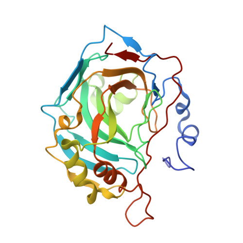
5EKH, 5EKJ, 5EKM - PubMed Abstract:
A supramolecular strategy for detecting specific proteins in complex media by using hyperpolarized (129) Xe NMR is reported. A cucurbit[6]uril (CB[6])-based molecular relay was programmed for three sequential equilibrium conditions by designing a two-faced guest (TFG) that initially binds CB[6] and blocks the CB[6]-Xe interaction. The protein analyte recruits the TFG and frees CB[6] for Xe binding. TFGs containing CB[6]- and carbonic anhydrase II (CAII)-binding domains were synthesized in one or two steps. X-ray crystallography confirmed TFG binding to Zn(2+) in the deep CAII active-site cleft, which precludes simultaneous CB[6] binding. The molecular relay was reprogrammed to detect avidin by using a different TFG. Finally, Xe binding by CB[6] was detected in buffer and in E. coli cultures expressing CAII through ultrasensitive (129) Xe NMR spectroscopy.
- Department of Chemistry, University of Pennsylvania, 231 South 34th Street, Philadelphia, PA, 19104-6323, USA.
Organizational Affiliation: