5EV8

Crystal structure of the metallo-beta-lactamase IMP-1 in complex with the bisthiazolidine inhibitor D-CS319


Experimental Data Snapshot

  • Method: X-RAY DIFFRACTION
  • Resolution: 2.30 Å
  • R-Value Free: 
    0.220 (Depositor), 0.220 (DCC) 
  • R-Value Work: 
    0.178 (Depositor), 0.180 (DCC) 
  • R-Value Observed: 
    0.180 (Depositor) 

Starting Model: experimental
View more details

wwPDB Validation   3D Report Full Report


Ligand Structure Quality Assessment 

Created with Raphaël 2.3.0Worse 01 BetterLigand structure goodness of fit to experimental dataBest fitted 3R9Click on this verticalbar to view details

This is version 1.3 of the entry. See complete history


Literature

Cross-class metallo-beta-lactamase inhibition by bisthiazolidines reveals multiple binding modes.

Hinchliffe, P.Gonzalez, M.M.Mojica, M.F.Gonzalez, J.M.Castillo, V.Saiz, C.Kosmopoulou, M.Tooke, C.L.Llarrull, L.I.Mahler, G.Bonomo, R.A.Vila, A.J.Spencer, J.

(2016) Proc Natl Acad Sci U S A 113: E3745-E3754

  • DOI: https://doi.org/10.1073/pnas.1601368113
  • Primary Citation of Related Structures:  
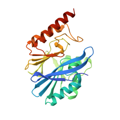
    4NQ6, 5EV6, 5EV8, 5EVB, 5EVD, 5EVK, 5EW0, 5EWA

  • PubMed Abstract: 

    Metallo-β-lactamases (MBLs) hydrolyze almost all β-lactam antibiotics and are unaffected by clinically available β-lactamase inhibitors (βLIs). Active-site architecture divides MBLs into three classes (B1, B2, and B3), complicating development of βLIs effective against all enzymes. Bisthiazolidines (BTZs) are carboxylate-containing, bicyclic compounds, considered as penicillin analogs with an additional free thiol. Here, we show both l- and d-BTZ enantiomers are micromolar competitive βLIs of all MBL classes in vitro, with Kis of 6-15 µM or 36-84 µM for subclass B1 MBLs (IMP-1 and BcII, respectively), and 10-12 µM for the B3 enzyme L1. Against the B2 MBL Sfh-I, the l-BTZ enantiomers exhibit 100-fold lower Kis (0.26-0.36 µM) than d-BTZs (26-29 µM). Importantly, cell-based time-kill assays show BTZs restore β-lactam susceptibility of Escherichia coli-producing MBLs (IMP-1, Sfh-1, BcII, and GOB-18) and, significantly, an extensively drug-resistant Stenotrophomonas maltophilia clinical isolate expressing L1. BTZs therefore inhibit the full range of MBLs and potentiate β-lactam activity against producer pathogens. X-ray crystal structures reveal insights into diverse BTZ binding modes, varying with orientation of the carboxylate and thiol moieties. BTZs bind the di-zinc centers of B1 (IMP-1; BcII) and B3 (L1) MBLs via the free thiol, but orient differently depending upon stereochemistry. In contrast, the l-BTZ carboxylate dominates interactions with the monozinc B2 MBL Sfh-I, with the thiol uninvolved. d-BTZ complexes most closely resemble β-lactam binding to B1 MBLs, but feature an unprecedented disruption of the D120-zinc interaction. Cross-class MBL inhibition therefore arises from the unexpected versatility of BTZ binding.


  • Organizational Affiliation

    School of Cellular and Molecular Medicine, Biomedical Sciences Building, University of Bristol, Bristol BS8 1TD, United Kingdom;


Macromolecules
Find similar proteins by:  (by identity cutoff)  |  3D Structure
Entity ID: 1
MoleculeChains Sequence LengthOrganismDetailsImage
Beta-lactamase IMP-1
A, B, C, D
230Serratia marcescensMutation(s): 0 
EC: 3.5.2.6 (PDB Primary Data), 3.1.26 (UniProt)
UniProt
Find proteins for P52699 (Serratia marcescens)
Explore P52699 
Go to UniProtKB:  P52699
Entity Groups  
Sequence Clusters30% Identity50% Identity70% Identity90% Identity95% Identity100% Identity
UniProt GroupP52699
Sequence Annotations
Expand
  • Reference Sequence
Small Molecules
Ligands 6 Unique
IDChains Name / Formula / InChI Key2D Diagram3D Interactions
3R9
Query on 3R9

Download Ideal Coordinates CCD File 
G [auth A],
H [auth A],
K [auth B],
R [auth C],
Y [auth D]
(3S,5S,7aR)-5-(sulfanylmethyl)tetrahydro[1,3]thiazolo[4,3-b][1,3]thiazole-3-carboxylic acid
C7 H11 N O2 S3
ZTWVMVSSSBGFHH-NGJCXOISSA-N
DMS
Query on DMS

Download Ideal Coordinates CCD File 
O [auth B],
T [auth C]
DIMETHYL SULFOXIDE
C2 H6 O S
IAZDPXIOMUYVGZ-UHFFFAOYSA-N
ZN
Query on ZN

Download Ideal Coordinates CCD File 
E [auth A]
F [auth A]
I [auth B]
J [auth B]
P [auth C]
ZINC ION
Zn
PTFCDOFLOPIGGS-UHFFFAOYSA-N
EDO
Query on EDO

Download Ideal Coordinates CCD File 
L [auth B],
M [auth B],
N [auth B],
S [auth C],
Z [auth D]
1,2-ETHANEDIOL
C2 H6 O2
LYCAIKOWRPUZTN-UHFFFAOYSA-N
CL
Query on CL

Download Ideal Coordinates CCD File 
V [auth C]CHLORIDE ION
Cl
VEXZGXHMUGYJMC-UHFFFAOYSA-M
NA
Query on NA

Download Ideal Coordinates CCD File 
U [auth C]SODIUM ION
Na
FKNQFGJONOIPTF-UHFFFAOYSA-N
Experimental Data & Validation

Experimental Data

  • Method: X-RAY DIFFRACTION
  • Resolution: 2.30 Å
  • R-Value Free:  0.220 (Depositor), 0.220 (DCC) 
  • R-Value Work:  0.178 (Depositor), 0.180 (DCC) 
  • R-Value Observed: 0.180 (Depositor) 
Space Group: P 21 21 21
Unit Cell:
Length ( Å )Angle ( ˚ )
a = 48.56α = 90
b = 77.713β = 90
c = 261.638γ = 90
Software Package:
Software NamePurpose
PHENIXrefinement
XDSdata reduction
Aimlessdata scaling
PHASERphasing

Structure Validation

View Full Validation Report


Ligand Structure Quality Assessment 

Created with Raphaël 2.3.0Worse 01 BetterLigand structure goodness of fit to experimental dataBest fitted 3R9Click on this verticalbar to view details

Entry History & Funding Information

Deposition Data


Funding OrganizationLocationGrant Number
National Institutes of Health/National Institute Of Allergy and Infectious Diseases (NIH/NIAID)United StatesR01AI100560

Revision History  (Full details and data files)

  • Version 1.0: 2016-06-01
    Type: Initial release
  • Version 1.1: 2016-07-20
    Changes: Database references
  • Version 1.2: 2022-03-30
    Changes: Author supporting evidence, Database references, Derived calculations
  • Version 1.3: 2024-01-10
    Changes: Data collection, Refinement description