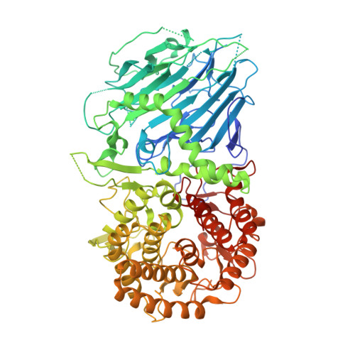
5FJS

Bacterial beta-glucosidase reveals the structural and functional basis of genetic defects in human glucocerebrosidase 2 (GBA2)


Experimental Data Snapshot

  • Method: X-RAY DIFFRACTION
  • Resolution: 2.60 Å
  • R-Value Free: 0.266 
  • R-Value Work: 0.206 
  • R-Value Observed: 0.209 

Starting Model: experimental
View more details

wwPDB Validation   3D Report Full Report


This is version 1.2 of the entry. See complete history


Literature

Bacterial Beta-Glucosidase Reveals the Structural and Functional Basis of Genetic Defects in Human Glucocerebrosidase 2 (Gba2)

Charoenwattanasatien, R.Pengthaisong, S.Breen, I.Mutoha, R.Sansenya, S.Hua, Y.Tankrathok, A.Wu, L.Songsiriritthigul, C.Tanaka, H.Williams, S.J.Davies, G.J.Kurisu, G.Ketudat Cairns, J.R.

(2016) ACS Chem Biol 11: 1891

  • DOI: https://doi.org/10.1021/acschembio.6b00192
  • Primary Citation of Related Structures:  
    5BVU, 5BX2, 5BX3, 5BX4, 5BX5, 5FJS

  • PubMed Abstract: 

    Human glucosylcerebrosidase 2 (GBA2) of the CAZy family GH116 is responsible for the breakdown of glycosphingolipids on the cytoplasmic face of the endoplasmic reticulum and Golgi apparatus. Genetic defects in GBA2 result in spastic paraplegia and cerebellar ataxia, while cross-talk between GBA2 and GBA1 glucosylceramidases may affect Gaucher disease. Here, we report the first three-dimensional structure for any GH116 enzyme, Thermoanaerobacterium xylanolyticum TxGH116 β-glucosidase, alone and in complex with diverse ligands. These structures allow identification of the glucoside binding and active site residues, which are shown to be conserved with GBA2. Mutagenic analysis of TxGH116 and structural modeling of GBA2 provide a detailed structural and functional rationale for pathogenic missense mutations of GBA2.


  • Organizational Affiliation

    Institute for Protein Research, Osaka University , 3-2 Yamadaoka, Suita, Osaka 565-0871, Japan.


Macromolecules
Find similar proteins by:  (by identity cutoff)  |  3D Structure
Entity ID: 1
MoleculeChains Sequence LengthOrganismDetailsImage
GLUCOSYLCERAMIDASE
A, B
787Thermoanaerobacterium xylanolyticum LX-11Mutation(s): 0 
EC: 3.2.1.21 (PDB Primary Data), 3.2.1.45 (UniProt)
UniProt
Find proteins for F6BL85 (Thermoanaerobacterium xylanolyticum (strain ATCC 49914 / DSM 7097 / LX-11))
Explore F6BL85 
Go to UniProtKB:  F6BL85
Entity Groups  
Sequence Clusters30% Identity50% Identity70% Identity90% Identity95% Identity100% Identity
UniProt GroupF6BL85
Sequence Annotations
Expand
  • Reference Sequence
Experimental Data & Validation

Experimental Data

  • Method: X-RAY DIFFRACTION
  • Resolution: 2.60 Å
  • R-Value Free: 0.266 
  • R-Value Work: 0.206 
  • R-Value Observed: 0.209 
  • Space Group: P 61
Unit Cell:
Length ( Å )Angle ( ˚ )
a = 187.974α = 90
b = 187.974β = 90
c = 99.075γ = 120
Software Package:
Software NamePurpose
REFMACrefinement
MOSFLMdata reduction
Aimlessdata scaling
MOLREPphasing

Structure Validation

View Full Validation Report


Entry History 

Revision History  (Full details and data files)

  • Version 1.0: 2016-05-11
    Type: Initial release
  • Version 1.1: 2016-07-27
    Changes: Database references
  • Version 1.2: 2024-01-10
    Changes: Data collection, Database references, Derived calculations, Other, Refinement description