5FTL

Cryo-EM structure of human p97 bound to ATPgS (Conformation I)


Experimental Data Snapshot

  • Method: ELECTRON MICROSCOPY
  • Resolution: 3.30 Å
  • Aggregation State: PARTICLE 
  • Reconstruction Method: SINGLE PARTICLE 

wwPDB Validation   3D Report Full Report


This is version 1.3 of the entry. See complete history


Literature

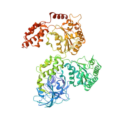
2.3 A Resolution Cryo-Em Structure of Human P97 and Mechanism of Allosteric Inhibition

Banerjee, S.Bartesaghi, A.Merk, A.Rao, P.Bulfer, S.L.Yan, Y.Green, N.Mroczkowski, B.Neitz, R.J.Wipf, P.Falconieri, V.Deshaies, R.J.Milne, J.L.S.Huryn, D.Arkin, M.Subramaniam, S.

(2016) Science 351: 871

  • DOI: https://doi.org/10.1126/science.aad7974
  • Primary Citation of Related Structures:  
    5FTJ, 5FTK, 5FTL, 5FTM, 5FTN

  • PubMed Abstract: 

    p97 is a hexameric AAA+ adenosine triphosphatase (ATPase) that is an attractive target for cancer drug development. We report cryo-electron microscopy (cryo-EM) structures for adenosine diphosphate (ADP)-bound, full-length, hexameric wild-type p97 in the presence and absence of an allosteric inhibitor at resolutions of 2.3 and 2.4 angstroms, respectively. We also report cryo-EM structures (at resolutions of ~3.3, 3.2, and 3.3 angstroms, respectively) for three distinct, coexisting functional states of p97 with occupancies of zero, one, or two molecules of adenosine 5'-O-(3-thiotriphosphate) (ATPγS) per protomer. A large corkscrew-like change in molecular architecture, coupled with upward displacement of the N-terminal domain, is observed only when ATPγS is bound to both the D1 and D2 domains of the protomer. These cryo-EM structures establish the sequence of nucleotide-driven structural changes in p97 at atomic resolution. They also enable elucidation of the binding mode of an allosteric small-molecule inhibitor to p97 and illustrate how inhibitor binding at the interface between the D1 and D2 domains prevents propagation of the conformational changes necessary for p97 function.


  • Organizational Affiliation

    Laboratory of Cell Biology, National Cancer Institute, Bethesda, MD 20892, USA.


Macromolecules
Find similar proteins by:  (by identity cutoff)  |  3D Structure
Entity ID: 1
MoleculeChains Sequence LengthOrganismDetailsImage
TRANSITIONAL ENDOPLASMIC RETICULUM ATPASE
A, B, C, D, E
A, B, C, D, E, F
806Homo sapiensMutation(s): 0 
EC: 3.6.4.6
UniProt & NIH Common Fund Data Resources
Find proteins for P55072 (Homo sapiens)
Explore P55072 
Go to UniProtKB:  P55072
PHAROS:  P55072
GTEx:  ENSG00000165280 
Entity Groups  
Sequence Clusters30% Identity50% Identity70% Identity90% Identity95% Identity100% Identity
UniProt GroupP55072
Sequence Annotations
Expand
  • Reference Sequence
Small Molecules
Ligands 1 Unique
IDChains Name / Formula / InChI Key2D Diagram3D Interactions
ADP
Query on ADP

Download Ideal Coordinates CCD File 
G [auth A]
H [auth A]
I [auth B]
J [auth B]
K [auth C]
G [auth A],
H [auth A],
I [auth B],
J [auth B],
K [auth C],
L [auth C],
M [auth D],
N [auth D],
O [auth E],
P [auth E],
Q [auth F],
R [auth F]
ADENOSINE-5'-DIPHOSPHATE
C10 H15 N5 O10 P2
XTWYTFMLZFPYCI-KQYNXXCUSA-N
Experimental Data & Validation

Experimental Data

  • Method: ELECTRON MICROSCOPY
  • Resolution: 3.30 Å
  • Aggregation State: PARTICLE 
  • Reconstruction Method: SINGLE PARTICLE 
EM Software:
TaskSoftware PackageVersion
RECONSTRUCTIONFREALIGN

Structure Validation

View Full Validation Report


Entry History 

Revision History  (Full details and data files)

  • Version 1.0: 2016-01-27
    Type: Initial release
  • Version 1.1: 2016-02-10
    Changes: Database references, Other
  • Version 1.2: 2016-03-09
    Changes: Database references
  • Version 1.3: 2018-10-03
    Changes: Data collection