5G0Q

beta-glucuronidase with an activity-based probe (N-alkyl cyclophellitol aziridine) bound


Experimental Data Snapshot

  • Method: X-RAY DIFFRACTION
  • Resolution: 1.65 Å
  • R-Value Free: 0.169 
  • R-Value Work: 0.120 
  • R-Value Observed: 0.123 

wwPDB Validation   3D Report Full Report


Ligand Structure Quality Assessment 


This is version 1.3 of the entry. See complete history


Literature

Activity-based probes for functional interrogation of retaining beta-glucuronidases.

Wu, L.Jiang, J.Jin, Y.Kallemeijn, W.W.Kuo, C.L.Artola, M.Dai, W.van Elk, C.van Eijk, M.van der Marel, G.A.Codee, J.D.C.Florea, B.I.Aerts, J.M.F.G.Overkleeft, H.S.Davies, G.J.

(2017) Nat Chem Biol 13: 867-873

  • DOI: https://doi.org/10.1038/nchembio.2395
  • Primary Citation of Related Structures:  
    5G0Q, 5L77, 5L9Y, 5L9Z, 5LA4, 5LA7

  • PubMed Abstract: 

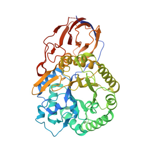
    Humans express at least two distinct β-glucuronidase enzymes that are involved in disease: exo-acting β-glucuronidase (GUSB), whose deficiency gives rise to mucopolysaccharidosis type VII, and endo-acting heparanase (HPSE), whose overexpression is implicated in inflammation and cancers. The medical importance of these enzymes necessitates reliable methods to assay their activities in tissues. Herein, we present a set of β-glucuronidase-specific activity-based probes (ABPs) that allow rapid and quantitative visualization of GUSB and HPSE in biological samples, providing a powerful tool for dissecting their activities in normal and disease states. Unexpectedly, we find that the supposedly inactive HPSE proenzyme proHPSE is also labeled by our ABPs, leading to surprising insights regarding structural relationships between proHPSE, mature HPSE, and their bacterial homologs. Our results demonstrate the application of β-glucuronidase ABPs in tracking pathologically relevant enzymes and provide a case study of how ABP-driven approaches can lead to discovery of unanticipated structural and biochemical functionality.


  • Organizational Affiliation

    York Structural Biology Laboratory, Department of Chemistry, University of York, Heslington, York, UK.


Macromolecules
Find similar proteins by:  (by identity cutoff)  |  3D Structure
Entity ID: 1
MoleculeChains Sequence LengthOrganismDetailsImage
BETA-GLUCURONIDASE475Acidobacterium capsulatumMutation(s): 0 
EC: 3.2.1.31
UniProt
Find proteins for C1F2K5 (Acidobacterium capsulatum (strain ATCC 51196 / DSM 11244 / BCRC 80197 / JCM 7670 / NBRC 15755 / NCIMB 13165 / 161))
Explore C1F2K5 
Go to UniProtKB:  C1F2K5
Entity Groups  
Sequence Clusters30% Identity50% Identity70% Identity90% Identity95% Identity100% Identity
UniProt GroupC1F2K5
Sequence Annotations
Expand
  • Reference Sequence
Experimental Data & Validation

Experimental Data

  • Method: X-RAY DIFFRACTION
  • Resolution: 1.65 Å
  • R-Value Free: 0.169 
  • R-Value Work: 0.120 
  • R-Value Observed: 0.123 
  • Space Group: C 1 2 1
Unit Cell:
Length ( Å )Angle ( ˚ )
a = 148.512α = 90
b = 44.896β = 114.78
c = 82.216γ = 90
Software Package:
Software NamePurpose
REFMACrefinement
xia2data reduction
xia2data scaling
MOLREPphasing

Structure Validation

View Full Validation Report


Ligand Structure Quality Assessment 


Entry History 

Deposition Data

Revision History  (Full details and data files)

  • Version 1.0: 2017-04-12
    Type: Initial release
  • Version 1.1: 2017-06-14
    Changes: Database references
  • Version 1.2: 2017-09-06
    Changes: Database references
  • Version 1.3: 2024-01-10
    Changes: Data collection, Database references, Derived calculations, Other, Refinement description