5G3N | pdb_00005g3n

Discovery of a novel secreted phospholipase A2 (sPLA2) inhibitor.


Experimental Data Snapshot

  • Method: X-RAY DIFFRACTION
  • Resolution: 1.80 Å
  • R-Value Free: 
    0.231 (Depositor), 0.232 (DCC) 
  • R-Value Work: 
    0.195 (Depositor), 0.193 (DCC) 
  • R-Value Observed: 
    0.197 (Depositor) 

wwPDB Validation 3D Report Full Report

Validation slider image for 5G3N

Ligand Structure Quality Assessment 


This is version 1.3 of the entry. See complete history

Literature

Discovery of Azd2716: A Novel Secreted Phospholipase A2 (Spla2) Inhibitor for the Treatment of Coronary Artery Disease

Giordanetto, F.Pettersen, D.Starke, I.Nordberg, P.Dahlstrom, M.Knerr, L.Selmi, N.Rosengren, B.Larsson, L.O.Sandmark, J.Castaldo, M.Dekker, N.Karlsson, U.Hurt-Camejo, E.

(2016) ACS Med Chem Lett 7: 884

  • DOI: https://doi.org/10.1021/acsmedchemlett.6b00188
  • Primary Citation Related Structures: 
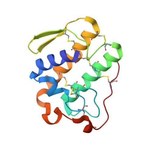
    5G3M, 5G3N

  • PubMed Abstract: 

    Expedited structure-based optimization of the initial fragment hit 1 led to the design of ( R )- 7 (AZD2716) a novel, potent secreted phospholipase A 2 (sPLA 2 ) inhibitor with excellent preclinical pharmacokinetic properties across species, clear in vivo efficacy, and minimized safety risk. Based on accumulated profiling data, ( R )- 7 was selected as a clinical candidate for the treatment of coronary artery disease.


  • Organizational Affiliation
    • Cardiovascular and Metabolic Diseases, Innovative Medicines and Early Development Biotech Unit Departments of Medicinal Chemistry, Bioscience, DMPK, Discovery Sciences Departments of Structure & Biophysics, Reagents and Assay Development, and Screening Sciences and Sample Management, Astrazeneca, Mölndal , Pepparedsleden 1, SE-431 83 Mölndal, Sweden.

Macromolecule Content 

  • Total Structure Weight: 29.41 kDa 
  • Atom Count: 2,238 
  • Modeled Residue Count: 252 
  • Deposited Residue Count: 254 
  • Unique protein chains: 1

Macromolecules

Find similar proteins by:|  3D Structure
Entity ID: 1
MoleculeChains  Sequence LengthOrganismDetailsImage
PHOSPHOLIPASE A2, MEMBRANE ASSOCIATED
A, B
127Homo sapiensMutation(s): 2 
EC: 3.1.1.4
UniProt & NIH Common Fund Data Resources
Find proteins for P14555 (Homo sapiens)
Explore P14555 
Go to UniProtKB:  P14555
PHAROS:  P14555
GTEx:  ENSG00000188257 
Entity Groups
Sequence Clusters30% Identity50% Identity70% Identity90% Identity95% Identity100% Identity
UniProt GroupP14555
Sequence Annotations
Expand
Reference Sequence

Small Molecules

Ligands 3 Unique
IDChains Name / Formula / InChI Key2D Diagram3D Interactions
X28

Query on X28


Download:Ideal Coordinates CCD File
F [auth A],
J [auth B]
3-(5'-BENZYL-2'-CARBAMOYLBIPHENYL-3-YL)PROPANOIC ACID
C23 H21 N O3
UHEGDWIZAOPGIY-UHFFFAOYSA-N
FMT

Query on FMT


Download:Ideal Coordinates CCD File
E [auth A],
I [auth B]
FORMIC ACID
C H2 O2
BDAGIHXWWSANSR-UHFFFAOYSA-N
CA

Query on CA


Download:Ideal Coordinates CCD File
C [auth A],
D [auth A],
G [auth B],
H [auth B]
CALCIUM ION
Ca
BHPQYMZQTOCNFJ-UHFFFAOYSA-N

Experimental Data & Validation

Experimental Data

  • Method: X-RAY DIFFRACTION
  • Resolution: 1.80 Å
  • R-Value Free:  0.231 (Depositor), 0.232 (DCC) 
  • R-Value Work:  0.195 (Depositor), 0.193 (DCC) 
  • R-Value Observed: 0.197 (Depositor) 
Space Group: P 21 21 21
Unit Cell:
Length ( Å )Angle ( ˚ )
a = 52.1α = 90
b = 66.53β = 90
c = 73.77γ = 90
Software Package:
Software NamePurpose
BUSTERrefinement
MOSFLMdata reduction
SCALAdata scaling
REFMACphasing

Structure Validation

View Full Validation Report


Ligand Structure Quality Assessment 


Entry History 

Deposition Data

Revision History  (Full details and data files)

  • Version 1.0: 2016-09-14
    Type: Initial release
  • Version 1.1: 2016-11-09
    Changes: Database references
  • Version 1.2: 2018-04-04
    Changes: Data collection
  • Version 1.3: 2024-11-06
    Changes: Data collection, Database references, Derived calculations, Other, Structure summary