5GLI

Human endothelin receptor type-B in the ligand-free form


Experimental Data Snapshot

  • Method: X-RAY DIFFRACTION
  • Resolution: 2.50 Å
  • R-Value Free: 
    0.276 (Depositor), 0.280 (DCC) 
  • R-Value Work: 
    0.247 (Depositor), 0.250 (DCC) 
  • R-Value Observed: 
    0.250 (Depositor) 

wwPDB Validation   3D Report Full Report


Ligand Structure Quality Assessment 

Created with Raphaël 2.3.0Worse 01 BetterLigand structure goodness of fit to experimental dataBest fitted OLCClick on this verticalbar to view detailsBest fitted OLAClick on this verticalbar to view details

This is version 1.4 of the entry. See complete history


Literature

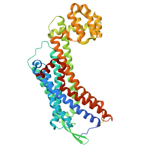
Activation mechanism of endothelin ETB receptor by endothelin-1.

Shihoya, W.Nishizawa, T.Okuta, A.Tani, K.Dohmae, N.Fujiyoshi, Y.Nureki, O.Doi, T.

(2016) Nature 537: 363-368

  • DOI: https://doi.org/10.1038/nature19319
  • Primary Citation of Related Structures:  
    5GLH, 5GLI

  • PubMed Abstract: 

    Endothelin, a 21-amino-acid peptide, participates in various physiological processes, such as regulation of vascular tone, humoral homeostasis, neural crest cell development and neurotransmission. Endothelin and its G-protein-coupled receptor are involved in the development of various diseases, such as pulmonary arterial hypertension, and thus are important therapeutic targets. Here we report crystal structures of human endothelin type B receptor in the ligand-free form and in complex with the endogenous agonist endothelin-1. The structures and mutation analysis reveal the mechanism for the isopeptide selectivity between endothelin-1 and -3. Transmembrane helices 1, 2, 6 and 7 move and envelop the entire endothelin peptide, in a virtually irreversible manner. The agonist-induced conformational changes are propagated to the receptor core and the cytoplasmic G-protein coupling interface, and probably induce conformational flexibility in TM6. A comparison with the M2 muscarinic receptor suggests a shared mechanism for signal transduction in class A G-protein-coupled receptors.


  • Organizational Affiliation

    Department of Basic Medicinal Sciences, Graduate School of Pharmaceutical Sciences, Nagoya University, Chikusa, Nagoya 464-8601, Japan.


Macromolecules
Find similar proteins by:  (by identity cutoff)  |  3D Structure
Entity ID: 1
MoleculeChains Sequence LengthOrganismDetailsImage
Endothelin Receptor Subtype-B464Homo sapiensMutation(s): 0 
Gene Names: EDNRB
EC: 3.2.1.17
Membrane Entity: Yes 
UniProt & NIH Common Fund Data Resources
Find proteins for P00720 (Enterobacteria phage T4)
Explore P00720 
Go to UniProtKB:  P00720
Find proteins for P24530 (Homo sapiens)
Explore P24530 
Go to UniProtKB:  P24530
PHAROS:  P24530
GTEx:  ENSG00000136160 
Entity Groups  
Sequence Clusters30% Identity50% Identity70% Identity90% Identity95% Identity100% Identity
UniProt GroupsP00720P24530
Sequence Annotations
Expand
  • Reference Sequence
Small Molecules
Ligands 3 Unique
IDChains Name / Formula / InChI Key2D Diagram3D Interactions
OLC
Query on OLC

Download Ideal Coordinates CCD File 
G [auth A],
I [auth A]
(2R)-2,3-dihydroxypropyl (9Z)-octadec-9-enoate
C21 H40 O4
RZRNAYUHWVFMIP-GDCKJWNLSA-N
OLA
Query on OLA

Download Ideal Coordinates CCD File 
H [auth A]OLEIC ACID
C18 H34 O2
ZQPPMHVWECSIRJ-KTKRTIGZSA-N
SO4
Query on SO4

Download Ideal Coordinates CCD File 
B [auth A],
C [auth A],
D [auth A],
E [auth A],
F [auth A]
SULFATE ION
O4 S
QAOWNCQODCNURD-UHFFFAOYSA-L
Experimental Data & Validation

Experimental Data

  • Method: X-RAY DIFFRACTION
  • Resolution: 2.50 Å
  • R-Value Free:  0.276 (Depositor), 0.280 (DCC) 
  • R-Value Work:  0.247 (Depositor), 0.250 (DCC) 
  • R-Value Observed: 0.250 (Depositor) 
Space Group: C 2 2 21
Unit Cell:
Length ( Å )Angle ( ˚ )
a = 73.98α = 90
b = 147.54β = 90
c = 107.81γ = 90
Software Package:
Software NamePurpose
PHENIXrefinement
PDB_EXTRACTdata extraction
XDSdata processing

Structure Validation

View Full Validation Report


Ligand Structure Quality Assessment 

Created with Raphaël 2.3.0Worse 01 BetterLigand structure goodness of fit to experimental dataBest fitted OLCClick on this verticalbar to view detailsBest fitted OLAClick on this verticalbar to view details

Entry History 

Deposition Data

Revision History  (Full details and data files)

  • Version 1.0: 2016-09-07
    Type: Initial release
  • Version 1.1: 2016-09-21
    Changes: Database references, Structure summary
  • Version 1.2: 2016-09-28
    Changes: Database references
  • Version 1.3: 2020-02-26
    Changes: Data collection, Database references, Derived calculations
  • Version 1.4: 2024-11-06
    Changes: Data collection, Database references, Structure summary