5H67

Crystal structure of the Bacillus subtilis SMC head domain complexed with the cognate ScpA C-terminal domain and soaked ATP


Experimental Data Snapshot

  • Method: X-RAY DIFFRACTION
  • Resolution: 2.07 Å
  • R-Value Free: 
    0.222 (Depositor), 0.220 (DCC) 
  • R-Value Work: 
    0.191 (Depositor), 0.190 (DCC) 
  • R-Value Observed: 
    0.192 (Depositor) 

Starting Model: experimental
View more details

wwPDB Validation   3D Report Full Report


Ligand Structure Quality Assessment 

Created with Raphaël 2.3.0Worse 01 BetterLigand structure goodness of fit to experimental dataBest fitted ATPClick on this verticalbar to view details

This is version 1.3 of the entry. See complete history


Literature

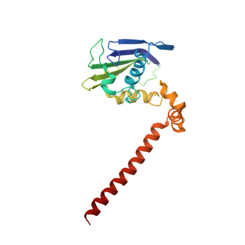
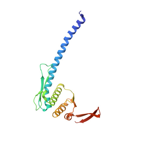
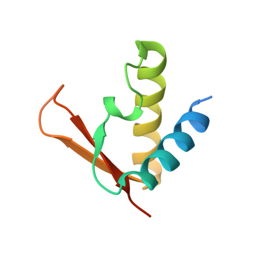
Overall Shapes of the SMC-ScpAB Complex Are Determined by Balance between Constraint and Relaxation of Its Structural Parts

Kamada, K.Su'etsugu, M.Takada, H.Miyata, M.Hirano, T.

(2017) Structure 25: 603-616.e4

  • DOI: https://doi.org/10.1016/j.str.2017.02.008
  • Primary Citation of Related Structures:  
    5H66, 5H67, 5H68, 5H69

  • PubMed Abstract: 

    The SMC-ScpAB complex plays a crucial role in chromosome organization and segregation in many bacteria. It is composed of a V-shaped SMC dimer and an ScpAB subcomplex that bridges the two Structural Maintenance of Chromosomes (SMC) head domains. Despite its functional significance, the mechanistic details of SMC-ScpAB remain obscure. Here we provide evidence that ATP-dependent head-head engagement induces a lever movement of the SMC neck region, which might help to separate juxtaposed coiled-coil arms. Binding of the ScpA N-terminal domain (NTD) to the SMC neck region is negatively regulated by the ScpB C-terminal domain. Mutations in the ScpA NTD compromise this regulation and profoundly affect the overall shape of the complex. The SMC hinge domain is structurally relaxed when free from coiled-coil juxtaposition. Taken together, we propose that the structural parts of SMC-ScpAB are subjected to the balance between constraint and relaxation, cooperating to modulate dynamic conformational changes of the whole complex.


  • Organizational Affiliation

    Chromosome Dynamics Laboratory, RIKEN, 2-1 Hirosawa, Wako, Saitama 351-0198, Japan. Electronic address: kamadak@riken.jp.


Macromolecules
Find similar proteins by:  (by identity cutoff)  |  3D Structure
Entity ID: 1
MoleculeChains Sequence LengthOrganismDetailsImage
Chromosome partition protein Smc199Bacillus subtilis subsp. subtilis str. 168Mutation(s): 0 
Gene Names: smcylqABSU15940
UniProt
Find proteins for P51834 (Bacillus subtilis (strain 168))
Explore P51834 
Go to UniProtKB:  P51834
Entity Groups  
Sequence Clusters30% Identity50% Identity70% Identity90% Identity95% Identity100% Identity
UniProt GroupP51834
Sequence Annotations
Expand
  • Reference Sequence
Find similar proteins by:  (by identity cutoff)  |  3D Structure
Entity ID: 2
MoleculeChains Sequence LengthOrganismDetailsImage
Chromosome partition protein Smc188Bacillus subtilis subsp. subtilis str. 168Mutation(s): 1 
Gene Names: smcylqABSU15940
UniProt
Find proteins for P51834 (Bacillus subtilis (strain 168))
Explore P51834 
Go to UniProtKB:  P51834
Entity Groups  
Sequence Clusters30% Identity50% Identity70% Identity90% Identity95% Identity100% Identity
UniProt GroupP51834
Sequence Annotations
Expand
  • Reference Sequence
Find similar proteins by:  (by identity cutoff)  |  3D Structure
Entity ID: 3
MoleculeChains Sequence LengthOrganismDetailsImage
Segregation and condensation protein A80Bacillus subtilis subsp. subtilis str. 168Mutation(s): 0 
Gene Names: scpAypuGBSU23220
UniProt
Find proteins for P35154 (Bacillus subtilis (strain 168))
Explore P35154 
Go to UniProtKB:  P35154
Entity Groups  
Sequence Clusters30% Identity50% Identity70% Identity90% Identity95% Identity100% Identity
UniProt GroupP35154
Sequence Annotations
Expand
  • Reference Sequence
Small Molecules
Ligands 1 Unique
IDChains Name / Formula / InChI Key2D Diagram3D Interactions
ATP
Query on ATP

Download Ideal Coordinates CCD File 
D [auth A]ADENOSINE-5'-TRIPHOSPHATE
C10 H16 N5 O13 P3
ZKHQWZAMYRWXGA-KQYNXXCUSA-N
Experimental Data & Validation

Experimental Data

  • Method: X-RAY DIFFRACTION
  • Resolution: 2.07 Å
  • R-Value Free:  0.222 (Depositor), 0.220 (DCC) 
  • R-Value Work:  0.191 (Depositor), 0.190 (DCC) 
  • R-Value Observed: 0.192 (Depositor) 
Space Group: P 1 21 1
Unit Cell:
Length ( Å )Angle ( ˚ )
a = 88.009α = 90
b = 47.124β = 112.594
c = 97.698γ = 90
Software Package:
Software NamePurpose
HKL-2000data reduction
SCALEPACKdata scaling
PHASERphasing
PHENIXrefinement

Structure Validation

View Full Validation Report


Ligand Structure Quality Assessment 

Created with Raphaël 2.3.0Worse 01 BetterLigand structure goodness of fit to experimental dataBest fitted ATPClick on this verticalbar to view details

Entry History & Funding Information

Deposition Data


Funding OrganizationLocationGrant Number
Grants-in-Aid for Science ResearchJapan--

Revision History  (Full details and data files)

  • Version 1.0: 2017-03-15
    Type: Initial release
  • Version 1.1: 2017-04-19
    Changes: Database references
  • Version 1.2: 2020-02-26
    Changes: Data collection
  • Version 1.3: 2023-11-08
    Changes: Data collection, Database references, Refinement description