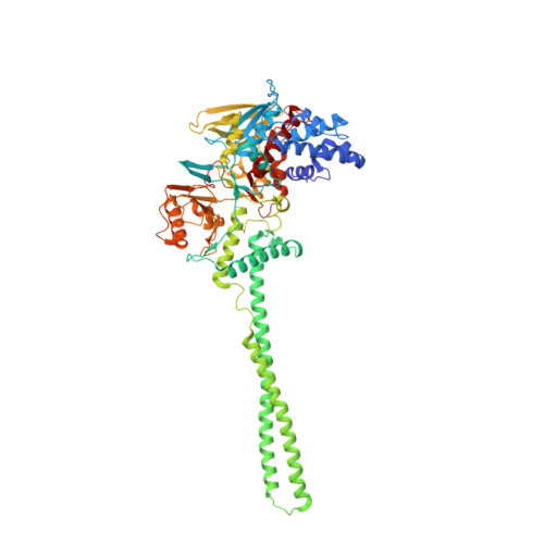
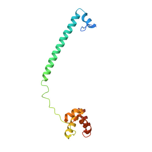
5H6R

Crystal structure of LSD1-CoREST in complex with peptide 13


Experimental Data Snapshot

  • Method: X-RAY DIFFRACTION
  • Resolution: 2.60 Å
  • R-Value Free: 0.231 
  • R-Value Work: 0.217 
  • R-Value Observed: 0.217 

Starting Model: experimental
View more details

wwPDB Validation   3D Report Full Report


Ligand Structure Quality Assessment 


This is version 2.0 of the entry. See complete history


Literature

Development and crystallographic evaluation of histone H3 peptide with N-terminal serine substitution as a potent inhibitor of lysine-specific demethylase 1.

Amano, Y.Kikuchi, M.Sato, S.Yokoyama, S.Umehara, T.Umezawa, N.Higuchi, T.

(2017) Bioorg Med Chem 25: 2617-2624

  • DOI: https://doi.org/10.1016/j.bmc.2017.03.016
  • Primary Citation of Related Structures:  
    5H6Q, 5H6R, 5X60

  • PubMed Abstract: 

    Lysine-specific demethylase 1 (LSD1/KDM1A) is a flavoenzyme demethylase, which removes mono- and dimethyl groups from histone H3 Lys4 (H3K4) or Lys9 (H3K9) in complexes with several nuclear proteins. Since LSD1 is implicated in the tumorigenesis and progression of various cancers, LSD1-specific inhibitors are considered as potential anti-cancer agents. A modified H3 peptide with substitution of Lys4 to Met [H3K4M] is already known to be a potent competitive inhibitor of LSD1. In this study, we synthesized a series of H3K4M peptide derivatives and evaluated their LSD1-inhibitory activities in vitro. We found that substitutions of the N-terminal amino acid with amino acids having a larger side chain were generally not tolerated, but substitution of Ala1 to Ser unexpectedly resulted in more potent inhibitory activity toward LSD1. X-ray crystallographic analysis of H3K4M derivatives bound to the LSD1·CoREST complex revealed the presence of additional hydrogen bonding between the N-terminal Ser residue of the H3 peptide derivative and LSD1. The present structural and biochemical findings will be helpful for obtaining more potent peptidic inhibitors of LSD1.


  • Organizational Affiliation

    Graduate School of Pharmaceutical Sciences, Nagoya City University, 3-1 Tanabe-dori, Mizuho-ku, Nagoya 467-8603, Japan.


Macromolecules
Find similar proteins by:  (by identity cutoff)  |  3D Structure
Entity ID: 1
MoleculeChains Sequence LengthOrganismDetailsImage
Lysine-specific histone demethylase 1A669Homo sapiensMutation(s): 0 
Gene Names: KDM1AAOF2KDM1KIAA0601LSD1
EC: 1 (PDB Primary Data), 1.14.99.66 (UniProt)
UniProt & NIH Common Fund Data Resources
Find proteins for O60341 (Homo sapiens)
Explore O60341 
Go to UniProtKB:  O60341
PHAROS:  O60341
GTEx:  ENSG00000004487 
Entity Groups  
Sequence Clusters30% Identity50% Identity70% Identity90% Identity95% Identity100% Identity
UniProt GroupO60341
Sequence Annotations
Expand
  • Reference Sequence
Find similar proteins by:  (by identity cutoff)  |  3D Structure
Entity ID: 2
MoleculeChains Sequence LengthOrganismDetailsImage
REST corepressor 1140Homo sapiensMutation(s): 0 
Gene Names: RCOR1KIAA0071RCOR
UniProt & NIH Common Fund Data Resources
Find proteins for Q9UKL0 (Homo sapiens)
Explore Q9UKL0 
Go to UniProtKB:  Q9UKL0
PHAROS:  Q9UKL0
GTEx:  ENSG00000089902 
Entity Groups  
Sequence Clusters30% Identity50% Identity70% Identity90% Identity95% Identity100% Identity
UniProt GroupQ9UKL0
Sequence Annotations
Expand
  • Reference Sequence

Find similar proteins by:  Sequence   |   3D Structure  

Entity ID: 3
MoleculeChains Sequence LengthOrganismDetailsImage
PEPTIDE (HSE)RTMQTARKSTGGKAPRKQLK21Homo sapiensMutation(s): 0 
UniProt & NIH Common Fund Data Resources
Find proteins for P68431 (Homo sapiens)
Explore P68431 
Go to UniProtKB:  P68431
PHAROS:  P68431
Entity Groups  
Sequence Clusters30% Identity50% Identity70% Identity90% Identity95% Identity100% Identity
UniProt GroupP68431
Sequence Annotations
Expand
  • Reference Sequence
Experimental Data & Validation

Experimental Data

  • Method: X-RAY DIFFRACTION
  • Resolution: 2.60 Å
  • R-Value Free: 0.231 
  • R-Value Work: 0.217 
  • R-Value Observed: 0.217 
  • Space Group: I 2 2 2
Unit Cell:
Length ( Å )Angle ( ˚ )
a = 121.674α = 90
b = 178.374β = 90
c = 234.848γ = 90
Software Package:
Software NamePurpose
REFMACrefinement
HKL-2000data processing
MOLREPphasing

Structure Validation

View Full Validation Report


Ligand Structure Quality Assessment 


Entry History 

Deposition Data

Revision History  (Full details and data files)

  • Version 1.0: 2017-04-12
    Type: Initial release
  • Version 1.1: 2017-12-06
    Changes: Data collection, Database references
  • Version 1.2: 2023-11-08
    Changes: Data collection, Database references, Refinement description
  • Version 2.0: 2023-11-15
    Changes: Atomic model, Data collection, Derived calculations