5HDH | pdb_00005hdh

Crystal structure of human TLR8 with an uncleaved Z-loop


Experimental Data Snapshot

  • Method: X-RAY DIFFRACTION
  • Resolution: 2.60 Å
  • R-Value Free: 
    0.239 (Depositor), 0.243 (DCC) 
  • R-Value Work: 
    0.206 (Depositor), 0.212 (DCC) 
  • R-Value Observed: 
    0.208 (Depositor) 

wwPDB Validation 3D Report Full Report

Validation slider image for 5HDH

Ligand Structure Quality Assessment 


This is version 2.1 of the entry. See complete history

Literature

Autoinhibition and relief mechanism by the proteolytic processing of Toll-like receptor 8

Tanji, H.Ohto, U.Motoi, Y.Shibata, T.Miyake, K.Shimizu, T.

(2016) Proc Natl Acad Sci U S A 113: 3012-3017

  • DOI: https://doi.org/10.1073/pnas.1516000113
  • Primary Citation Related Structures: 
    5HDH

  • PubMed Abstract: 

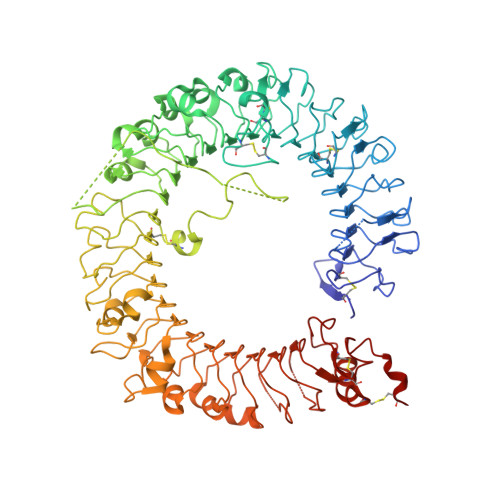
    Toll-like receptor 8 (TLR8) senses single-stranded RNA (ssRNA) and initiates innate immune responses. TLR8 requires proteolytic cleavage at the loop region (Z-loop) between leucine-rich repeat (LRR) 14 and LRR15 for its activation. However, the molecular basis of Z-loop processing remains unknown. To elucidate the mechanism of Z-loop processing, we performed biochemical and structural studies of how the Z-loop affects the function of TLR8. TLR8 with the uncleaved Z-loop is unable to form a dimer, which is essential for activation, irrespective of the presence of agonistic ligands. Crystallographic analysis revealed that the uncleaved Z-loop located on the ascending lateral face prevents the approach of the dimerization partner by steric hindrance. This autoinhibition mechanism of dimerization by the Z-loop might be occurring in the proteins of the same subfamily, TLR7 and TLR9.


  • Organizational Affiliation
    • Graduate School of Pharmaceutical Sciences, The University of Tokyo, Tokyo 113-0033, Japan;

Macromolecule Content 

  • Total Structure Weight: 95.3 kDa 
  • Atom Count: 6,371 
  • Modeled Residue Count: 751 
  • Deposited Residue Count: 801 
  • Unique protein chains: 1

Macromolecules

Find similar proteins by:|  3D Structure
Entity ID: 1
MoleculeChains  Sequence LengthOrganismDetailsImage
Toll-like receptor 8801Homo sapiensMutation(s): 4 
Gene Names: TLR8UNQ249/PRO286
UniProt & NIH Common Fund Data Resources
Find proteins for Q9NR97 (Homo sapiens)
Explore Q9NR97 
Go to UniProtKB:  Q9NR97
PHAROS:  Q9NR97
GTEx:  ENSG00000101916 
Entity Groups
Sequence Clusters30% Identity50% Identity70% Identity90% Identity95% Identity100% Identity
UniProt GroupQ9NR97
Glycosylation
Glycosylation Sites: 9Go to GlyGen: Q9NR97-1
Sequence Annotations
Expand
Reference Sequence

Oligosaccharides

Help  
Entity ID: 2
MoleculeChains Length2D Diagram GlycosylationD Interactions
alpha-D-mannopyranose-(1-6)-beta-D-mannopyranose-(1-4)-2-acetamido-2-deoxy-beta-D-glucopyranose-(1-4)-2-acetamido-2-deoxy-beta-D-glucopyranose
B
4N-Glycosylation
Glycosylation Resources
GlyTouCan: G22573RC
GlyCosmos: G22573RC
GlyGen: G22573RC
Entity ID: 3
MoleculeChains Length2D Diagram GlycosylationD Interactions
2-acetamido-2-deoxy-beta-D-glucopyranose-(1-4)-2-acetamido-2-deoxy-beta-D-glucopyranose
C
2N-Glycosylation
Glycosylation Resources
GlyTouCan: G42666HT
GlyCosmos: G42666HT
GlyGen: G42666HT
Entity ID: 4
MoleculeChains Length2D Diagram GlycosylationD Interactions
alpha-D-mannopyranose-(1-3)-[alpha-D-mannopyranose-(1-6)]beta-D-mannopyranose-(1-4)-2-acetamido-2-deoxy-beta-D-glucopyranose-(1-4)-2-acetamido-2-deoxy-beta-D-glucopyranose
D
5N-Glycosylation
Glycosylation Resources
GlyTouCan: G22768VO
GlyCosmos: G22768VO
GlyGen: G22768VO

Small Molecules

Ligands 3 Unique
IDChains Name / Formula / InChI Key2D Diagram3D Interactions
NAG

Query on NAG


Download:Ideal Coordinates CCD File
E [auth A]
F [auth A]
G [auth A]
H [auth A]
I [auth A]
E [auth A],
F [auth A],
G [auth A],
H [auth A],
I [auth A],
J [auth A]
2-acetamido-2-deoxy-beta-D-glucopyranose
C8 H15 N O6
OVRNDRQMDRJTHS-FMDGEEDCSA-N
MES

Query on MES


Download:Ideal Coordinates CCD File
N [auth A]2-(N-MORPHOLINO)-ETHANESULFONIC ACID
C6 H13 N O4 S
SXGZJKUKBWWHRA-UHFFFAOYSA-N
SO4

Query on SO4


Download:Ideal Coordinates CCD File
K [auth A],
L [auth A],
M [auth A]
SULFATE ION
O4 S
QAOWNCQODCNURD-UHFFFAOYSA-L

Experimental Data & Validation

Experimental Data

  • Method: X-RAY DIFFRACTION
  • Resolution: 2.60 Å
  • R-Value Free:  0.239 (Depositor), 0.243 (DCC) 
  • R-Value Work:  0.206 (Depositor), 0.212 (DCC) 
  • R-Value Observed: 0.208 (Depositor) 
Space Group: H 3 2
Unit Cell:
Length ( Å )Angle ( ˚ )
a = 171.52α = 90
b = 171.52β = 90
c = 301.333γ = 120
Software Package:
Software NamePurpose
REFMACrefinement
XDSdata processing
Aimlessdata scaling
MOLREPphasing

Structure Validation

View Full Validation Report


Ligand Structure Quality Assessment 


Entry History 

Deposition Data

Revision History  (Full details and data files)

  • Version 1.0: 2016-03-09
    Type: Initial release
  • Version 1.1: 2016-06-22
    Changes: Other
  • Version 1.2: 2020-02-19
    Changes: Data collection, Derived calculations
  • Version 2.0: 2020-07-29
    Type: Remediation
    Reason: Carbohydrate remediation
    Changes: Atomic model, Data collection, Derived calculations, Structure summary
  • Version 2.1: 2024-10-30
    Changes: Data collection, Database references, Derived calculations, Structure summary