Identification of a 5-[3-phenyl-(2-cyclic-ether)-methylether]-4-aminopyrrolo[2,3-d]pyrimidine series of IGF-1R inhibitors.
Stauffer, F., Cowan-Jacob, S.W., Scheufler, C., Furet, P.(2016) Bioorg Med Chem Lett 26: 2065-2067
- PubMed: 26951750 Search on PubMed
- DOI: https://doi.org/10.1016/j.bmcl.2016.02.074
- Primary Citation Related Structures:
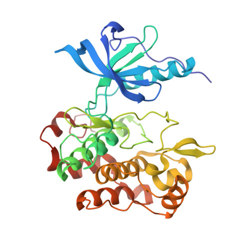
5HHW, 5HZN - PubMed Abstract:
We report structure-guided modifications of the benzyloxy substituent of the Insulin-like Growth Factor-1 Receptor (IGF-1R) inhibitor NVP-AEW541. This chemical group has been shown to confer selectivity against other protein kinases but at the expense of a metabolism liability. X-ray crystallography has revealed that the benzyloxy moiety interacts with a lysine cation of the IGF-1R kinase domain via its ether function and its aromatic π-system and is nicely embedded in an induced hydrophobic pocket. We show that 1,4-diethers displaying an adequate hydrophobic and constrained shape are advantageous benzyloxy replacements. A single digit nanomolar inhibitor (compound 20, IC50=8.9 nM) was identified following this approach.
- Novartis Institutes for Biomedical Research, Basel, Postfach, 4002 Basel, Switzerland. Electronic address: frederic.stauffer@novartis.com.
Organizational Affiliation: