Structural Basis for Eculizumab-Mediated Inhibition of the Complement Terminal Pathway.
Schatz-Jakobsen, J.A., Zhang, Y., Johnson, K., Neill, A., Sheridan, D., Andersen, G.R.(2016) J Immunol 197: 337-344
- PubMed: 27194791 Search on PubMed
- DOI: https://doi.org/10.4049/jimmunol.1600280
- Primary Citation Related Structures:
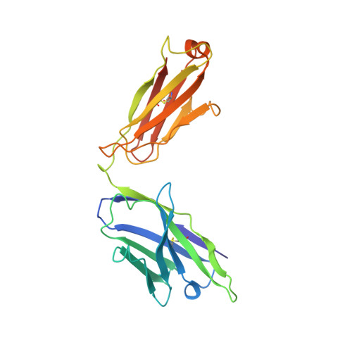
5I5K - PubMed Abstract:
Eculizumab is a humanized mAb approved for treatment of patients with paroxysmal nocturnal hemoglobinuria and atypical hemolytic uremic syndrome. Eculizumab binds complement component C5 and prevents its cleavage by C5 convertases, inhibiting release of both the proinflammatory metabolite C5a and formation of the membrane attack complex via C5b. In this study, we present the crystal structure of the complex between C5 and a Fab fragment with the same sequence as eculizumab at a resolution of 4.2 Å. Five CDRs contact the C5 macroglobulin 7 domain, which contains the entire epitope. A complete mutational scan of the 66 CDR residues identified 28 residues as important for the C5-eculizumab interaction, and the structure of the complex offered an explanation for the reduced C5 binding observed for these mutant Abs. Furthermore, the structural observations of the interaction are supported by the reduced ability of a subset of these mutated Abs to inhibit membrane attack complex formation as tested in a hemolysis assay. Our results suggest that eculizumab functions by sterically preventing C5 from binding to convertases and explain the exquisite selectivity of eculizumab for human C5 and how polymorphisms in C5 cause eculizumab-resistance in a small number of patients with paroxysmal nocturnal hemoglobinuria.
- Department of Molecular Biology and Genetics, Aarhus University, DK-8000 Aarhus, Denmark; and.
Organizational Affiliation: