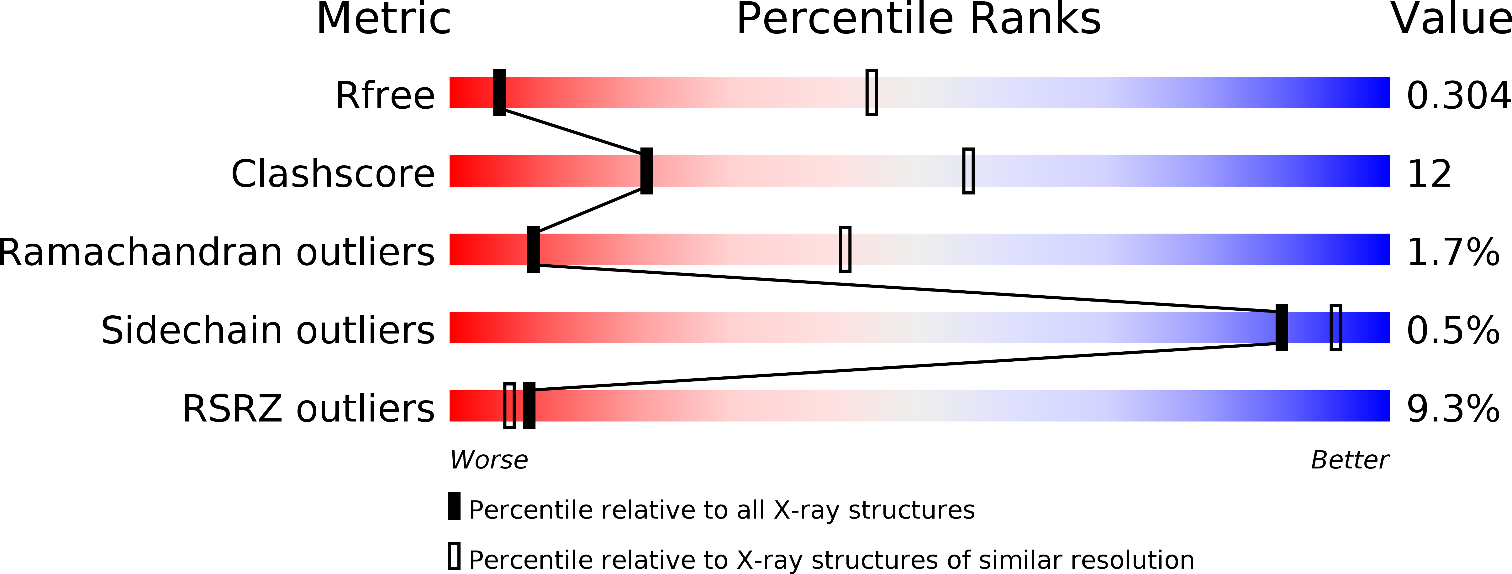
5I6C

The structure of the eukaryotic purine/H+ symporter, UapA, in complex with Xanthine


Experimental Data Snapshot

  • Method: X-RAY DIFFRACTION
  • Resolution: 3.70 Å
  • R-Value Free: 
    0.327 (Depositor), 0.300 (DCC) 
  • R-Value Work: 
    0.296 (Depositor), 0.300 (DCC) 
  • R-Value Observed: 
    0.298 (Depositor) 

wwPDB Validation   3D Report Full Report


Ligand Structure Quality Assessment 

Created with Raphaël 2.3.0Worse 01 BetterLigand structure goodness of fit to experimental dataBest fitted LMTClick on this verticalbar to view detailsBest fitted XANClick on this verticalbar to view details

This is version 1.3 of the entry. See complete history


Literature

Structure of eukaryotic purine/H(+) symporter UapA suggests a role for homodimerization in transport activity.

Alguel, Y.Amillis, S.Leung, J.Lambrinidis, G.Capaldi, S.Scull, N.J.Craven, G.Iwata, S.Armstrong, A.Mikros, E.Diallinas, G.Cameron, A.D.Byrne, B.

(2016) Nat Commun 7: 11336-11336

  • DOI: https://doi.org/10.1038/ncomms11336
  • Primary Citation of Related Structures:  
    5I6C

  • PubMed Abstract: 

    The uric acid/xanthine H(+) symporter, UapA, is a high-affinity purine transporter from the filamentous fungus Aspergillus nidulans. Here we present the crystal structure of a genetically stabilized version of UapA (UapA-G411VΔ1-11) in complex with xanthine. UapA is formed from two domains, a core domain and a gate domain, similar to the previously solved uracil transporter UraA, which belongs to the same family. The structure shows UapA in an inward-facing conformation with xanthine bound to residues in the core domain. Unlike UraA, which was observed to be a monomer, UapA forms a dimer in the crystals with dimer interactions formed exclusively through the gate domain. Analysis of dominant negative mutants is consistent with dimerization playing a key role in transport. We postulate that UapA uses an elevator transport mechanism likely to be shared with other structurally homologous transporters including anion exchangers and prestin.


  • Organizational Affiliation

    Department of Life Sciences, Imperial College London, London SW7 2AZ, UK.


Macromolecules
Find similar proteins by:  (by identity cutoff)  |  3D Structure
Entity ID: 1
MoleculeChains Sequence LengthOrganismDetailsImage
Uric acid-xanthine permease
A, B
574Aspergillus nidulans FGSC A4Mutation(s): 1 
Gene Names: uapAAN6932
Membrane Entity: Yes 
UniProt
Find proteins for Q07307 (Emericella nidulans (strain FGSC A4 / ATCC 38163 / CBS 112.46 / NRRL 194 / M139))
Explore Q07307 
Go to UniProtKB:  Q07307
Entity Groups  
Sequence Clusters30% Identity50% Identity70% Identity90% Identity95% Identity100% Identity
UniProt GroupQ07307
Sequence Annotations
Expand
  • Reference Sequence
Experimental Data & Validation

Experimental Data

  • Method: X-RAY DIFFRACTION
  • Resolution: 3.70 Å
  • R-Value Free:  0.327 (Depositor), 0.300 (DCC) 
  • R-Value Work:  0.296 (Depositor), 0.300 (DCC) 
  • R-Value Observed: 0.298 (Depositor) 
Space Group: P 1 21 1
Unit Cell:
Length ( Å )Angle ( ˚ )
a = 75.09α = 90
b = 173.83β = 111.11
c = 82.39γ = 90
Software Package:
Software NamePurpose
PHENIXrefinement
xia2data reduction
xia2data scaling
SHARPphasing

Structure Validation

View Full Validation Report


Ligand Structure Quality Assessment 

Created with Raphaël 2.3.0Worse 01 BetterLigand structure goodness of fit to experimental dataBest fitted LMTClick on this verticalbar to view detailsBest fitted XANClick on this verticalbar to view details

Entry History & Funding Information

Deposition Data


Funding OrganizationLocationGrant Number
Biotechnology and Biological Sciences Research CouncilUnited Kingdom--

Revision History  (Full details and data files)

  • Version 1.0: 2016-04-27
    Type: Initial release
  • Version 1.1: 2016-05-04
    Changes: Database references
  • Version 1.2: 2017-08-30
    Changes: Author supporting evidence
  • Version 1.3: 2024-11-13
    Changes: Data collection, Database references, Structure summary