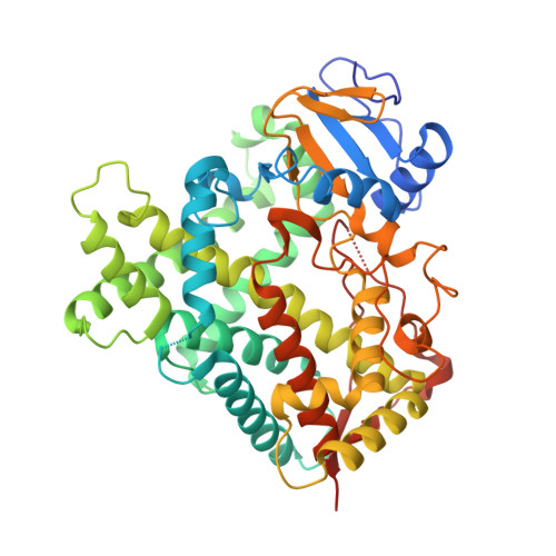
5IUT

STRUCTURE OF P450 2B4 F202W MUTANT


Experimental Data Snapshot

  • Method: X-RAY DIFFRACTION
  • Resolution: 2.34 Å
  • R-Value Free: 0.265 
  • R-Value Work: 0.216 
  • R-Value Observed: 0.218 

Starting Model: experimental
View more details

wwPDB Validation   3D Report Full Report


Ligand Structure Quality Assessment 


This is version 1.4 of the entry. See complete history


Literature

Effect of detergent binding on cytochrome P450 2B4 structure as analyzed by X-ray crystallography and deuterium-exchange mass spectrometry.

Shah, M.B.Jang, H.H.Wilderman, P.R.Lee, D.Li, S.Zhang, Q.Stout, C.D.Halpert, J.R.

(2016) Biophys Chem 216: 1-8

  • DOI: https://doi.org/10.1016/j.bpc.2016.05.007
  • Primary Citation of Related Structures:  
    5IUT, 5IUZ

  • PubMed Abstract: 

    Multiple crystal structures of CYP2B4 have demonstrated the binding of the detergent 5-cyclohexyl-1-pentyl-β-D-maltoside (CYMAL-5) in a peripheral pocket located adjacent to the active site. To explore the consequences of detergent binding, X-ray crystal structures of the peripheral pocket mutant CYP2B4 F202W were solved in the presence of hexaethylene glycol monooctyl ether (C8E6) and CYMAL-5. The structure in the presence of CYMAL-5 illustrated a closed conformation indistinguishable from the previously solved wild-type. In contrast, the F202W structure in the presence of C8E6 revealed a detergent molecule that coordinated the heme-iron and extended to the protein surface through the substrate access channel 2f. Despite the overall structural similarity of these detergent complexes, remarkable differences were observed in the A, A', and H helices, the F-G cassette, the C-D and β4 loop region. Hydrogen-deuterium exchange mass spectrometry (DXMS) was employed to probe these differences and to test the effect of detergents in solution. The presence of either detergent increased the H/D exchange rate across the plastic regions, and the results obtained by DXMS in solution were consistent in general with the relevant structural snapshots. The study provides insight into effect of detergent binding and the interpretation of associated conformational dynamics of CYP2B4.


  • Organizational Affiliation

    School of Pharmacy, University of Connecticut, Storrs, CT 06269, United States. Electronic address: manish.shah@uconn.edu.


Macromolecules
Find similar proteins by:  (by identity cutoff)  |  3D Structure
Entity ID: 1
MoleculeChains Sequence LengthOrganismDetailsImage
Cytochrome P450 2B4478Oryctolagus cuniculusMutation(s): 10 
Gene Names: CYP2B4
EC: 1.14.14.1
Membrane Entity: Yes 
UniProt
Find proteins for P00178 (Oryctolagus cuniculus)
Explore P00178 
Go to UniProtKB:  P00178
Entity Groups  
Sequence Clusters30% Identity50% Identity70% Identity90% Identity95% Identity100% Identity
UniProt GroupP00178
Sequence Annotations
Expand
  • Reference Sequence
Small Molecules
Ligands 2 Unique
IDChains Name / Formula / InChI Key2D Diagram3D Interactions
HEM
Query on HEM

Download Ideal Coordinates CCD File 
B [auth A]PROTOPORPHYRIN IX CONTAINING FE
C34 H32 Fe N4 O4
KABFMIBPWCXCRK-RGGAHWMASA-L
32M
Query on 32M

Download Ideal Coordinates CCD File 
C [auth A]3,6,9,12,15,18-hexaoxahexacosan-1-ol
C20 H42 O7
VVSOZSGWKFPDFX-UHFFFAOYSA-N
Experimental Data & Validation

Experimental Data

  • Method: X-RAY DIFFRACTION
  • Resolution: 2.34 Å
  • R-Value Free: 0.265 
  • R-Value Work: 0.216 
  • R-Value Observed: 0.218 
  • Space Group: P 31 2 1
Unit Cell:
Length ( Å )Angle ( ˚ )
a = 91.165α = 90
b = 91.165β = 90
c = 151.137γ = 120
Software Package:
Software NamePurpose
autoXDSdata reduction
SCALAdata scaling
PHASERphasing
REFMACrefinement

Structure Validation

View Full Validation Report


Ligand Structure Quality Assessment 


Entry History & Funding Information

Deposition Data


Funding OrganizationLocationGrant Number
National Institutes of Health/National Institute of Environmental Health Sciences (NIH/NIEHS)United StatesES003619

Revision History  (Full details and data files)

  • Version 1.0: 2016-06-08
    Type: Initial release
  • Version 1.1: 2016-06-22
    Changes: Database references
  • Version 1.2: 2017-09-20
    Changes: Author supporting evidence, Database references, Derived calculations
  • Version 1.3: 2019-12-18
    Changes: Author supporting evidence, Database references
  • Version 1.4: 2023-09-27
    Changes: Data collection, Database references, Refinement description