5J5Z | pdb_00005j5z

Crystal structure of the D444V disease-causing mutant of the human dihydrolipoamide dehydrogenase


Experimental Data Snapshot

  • Method: X-RAY DIFFRACTION
  • Resolution: 1.84 Å
  • R-Value Free: 
    0.226 (Depositor), 0.230 (DCC) 
  • R-Value Work: 
    0.181 (Depositor), 0.190 (DCC) 
  • R-Value Observed: 
    0.182 (Depositor) 

Starting Model: experimental
View more details

wwPDB Validation   3D Report Full Report


Ligand Structure Quality Assessment 

Created with Raphaël 2.3.0Worse 01 BetterLigand structure goodness of fit to experimental dataBest fitted FADClick on this verticalbar to view details

This is version 1.5 of the entry. See complete history


Literature

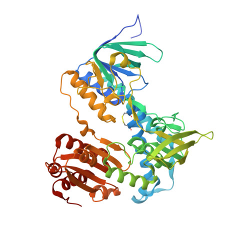
Crystal structures of the disease-causing D444V mutant and the relevant wild type human dihydrolipoamide dehydrogenase.

Szabo, E.Mizsei, R.Wilk, P.Zambo, Z.Torocsik, B.Weiss, M.S.Adam-Vizi, V.Ambrus, A.

(2018) Free Radic Biol Med 124: 214-220

  • DOI: https://doi.org/10.1016/j.freeradbiomed.2018.06.008
  • Primary Citation of Related Structures:  
    5J5Z, 5NHG

  • PubMed Abstract: 

    We report the crystal structures of the human (dihydro)lipoamide dehydrogenase (hLADH, hE3) and its disease-causing homodimer interface mutant D444V-hE3 at 2.27 and 1.84 Å resolution, respectively. The wild type structure is a unique uncomplexed, unliganded hE3 structure with the true canonical sequence. Based on the structural information a novel molecular pathomechanism is proposed for the impaired catalytic activity and enhanced capacity for reactive oxygen species generation of the pathogenic mutant. The mechanistic model involves a previously much ignored solvent accessible channel leading to the active site that might be perturbed also by other disease-causing homodimer interface substitutions of this enzyme.


  • Organizational Affiliation

    Department of Medical Biochemistry, MTA-SE Laboratory for Neurobiochemistry, Semmelweis University, H-1094 Budapest, Hungary.


Macromolecules
Find similar proteins by:  (by identity cutoff)  |  3D Structure
Entity ID: 1
MoleculeChains Sequence LengthOrganismDetailsImage
Dihydrolipoyl dehydrogenase, mitochondrial
A, B
496Homo sapiensMutation(s): 1 
Gene Names: DLDGCSLLADPHE3
EC: 1.8.1.4
UniProt & NIH Common Fund Data Resources
Find proteins for P09622 (Homo sapiens)
Explore P09622 
Go to UniProtKB:  P09622
PHAROS:  P09622
GTEx:  ENSG00000091140 
Entity Groups  
Sequence Clusters30% Identity50% Identity70% Identity90% Identity95% Identity100% Identity
UniProt GroupP09622
Sequence Annotations
Expand
  • Reference Sequence
Small Molecules
Ligands 3 Unique
IDChains Name / Formula / InChI Key2D Diagram3D Interactions
FAD
Query on FAD

Download Ideal Coordinates CCD File 
C [auth A],
H [auth B]
FLAVIN-ADENINE DINUCLEOTIDE
C27 H33 N9 O15 P2
VWWQXMAJTJZDQX-UYBVJOGSSA-N
PO4
Query on PO4

Download Ideal Coordinates CCD File 
D [auth A],
I [auth B]
PHOSPHATE ION
O4 P
NBIIXXVUZAFLBC-UHFFFAOYSA-K
GOL
Query on GOL

Download Ideal Coordinates CCD File 
E [auth A],
F [auth A],
G [auth A]
GLYCEROL
C3 H8 O3
PEDCQBHIVMGVHV-UHFFFAOYSA-N
Experimental Data & Validation

Experimental Data

  • Method: X-RAY DIFFRACTION
  • Resolution: 1.84 Å
  • R-Value Free:  0.226 (Depositor), 0.230 (DCC) 
  • R-Value Work:  0.181 (Depositor), 0.190 (DCC) 
  • R-Value Observed: 0.182 (Depositor) 
Space Group: P 21 21 2
Unit Cell:
Length ( Å )Angle ( ˚ )
a = 118.035α = 90
b = 168.939β = 90
c = 61.279γ = 90
Software Package:
Software NamePurpose
REFMACrefinement
MxCuBEdata collection
XDSdata reduction
XDSdata scaling
MOLREPphasing

Structure Validation

View Full Validation Report


Ligand Structure Quality Assessment 

Created with Raphaël 2.3.0Worse 01 BetterLigand structure goodness of fit to experimental dataBest fitted FADClick on this verticalbar to view details

Entry History & Funding Information

Deposition Data


Funding OrganizationLocationGrant Number
Hungarian Academy of Sciences (MTA)Hungary02001
Hungarian Scientific Research Fund (OTKA)Hungary112230
Hungarian Brain Research Program (NAP)HungaryKTIA_13_NAP_A_III/6
European Molecular Biology OrganizationHungaryShort-term Fellowship [to A.A.]
Hungarian Academy of SciencesHungaryBolyai Fellowship [to A.A]

Revision History  (Full details and data files)

  • Version 1.0: 2017-11-15
    Type: Initial release
  • Version 1.1: 2018-03-28
    Changes: Source and taxonomy
  • Version 1.2: 2018-07-04
    Changes: Data collection, Database references, Derived calculations
  • Version 1.3: 2018-07-11
    Changes: Data collection, Database references
  • Version 1.4: 2024-01-10
    Changes: Data collection, Database references, Refinement description
  • Version 1.5: 2024-10-16
    Changes: Structure summary