1,4,5-Trisubstituted Imidazole-Based p53-MDM2/MDMX Antagonists with Aliphatic Linkers for Conjugation with Biological Carriers.
Twarda-Clapa, A., Krzanik, S., Kubica, K., Guzik, K., Labuzek, B., Neochoritis, C.G., Khoury, K., Kowalska, K., Czub, M., Dubin, G., Domling, A., Skalniak, L., Holak, T.A.(2017) J Med Chem 60: 4234-4244
- PubMed: 28482147 Search on PubMed
- DOI: https://doi.org/10.1021/acs.jmedchem.7b00104
- Primary Citation Related Structures:
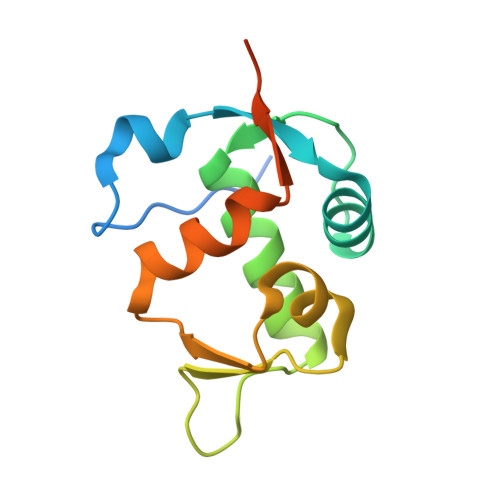
5J7F, 5J7G - PubMed Abstract:
The tumor suppressor protein p53, the "guardian of the genome", is inactivated in nearly all cancer types by mutations in the TP53 gene or by overexpression of its negative regulators, oncoproteins MDM2/MDMX. Recovery of p53 function by disrupting the p53-MDM2/MDMX interaction using small-molecule antagonists could provide an efficient nongenotoxic anticancer therapy. Here we present the syntheses, activities, and crystal structures of the p53-MDM2/MDMX inhibitors based on the 1,4,5-trisubstituted imidazole scaffold which are appended with aliphatic linkers that enable coupling to bioactive carriers. The compounds have favorable properties at both biochemical and cellular levels. The most effective compound 19 is a tight binder of MDM2 and activates p53 in cancer cells that express the wild-type p53, leading to cell cycle arrest and growth inhibition. Crystal structures reveal that compound 19 induces MDM2 dimerization via the aliphatic linker. This unique dimerization-binding mode opens new prospects for the optimization of the p53-MDM2/MDMX inhibitors and conjugation with bioactive carriers.
- Faculty of Biochemistry, Biophysics, and Biotechnology, Jagiellonian University , Gronostajowa 7, 30-387 Cracow, Poland.
Organizational Affiliation: