5JDU

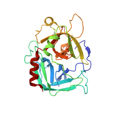
Crystal structure for human thrombin mutant D189A


Experimental Data Snapshot

  • Method: X-RAY DIFFRACTION
  • Resolution: 1.70 Å
  • R-Value Free: 0.206 
  • R-Value Work: 0.181 
  • R-Value Observed: 0.182 

Starting Model: experimental
View more details

wwPDB Validation   3D Report Full Report


This is version 2.2 of the entry. See complete history


Literature

Loop Electrostatics Asymmetry Modulates the Preexisting Conformational Equilibrium in Thrombin.

Pozzi, N.Zerbetto, M.Acquasaliente, L.Tescari, S.Frezzato, D.Polimeno, A.Gohara, D.W.Di Cera, E.De Filippis, V.

(2016) Biochemistry 55: 3984-3994

  • DOI: https://doi.org/10.1021/acs.biochem.6b00385
  • Primary Citation of Related Structures:  
    5JDU

  • PubMed Abstract: 

    Thrombin exists as an ensemble of active (E) and inactive (E*) conformations that differ in their accessibility to the active site. Here we show that redistribution of the E*-E equilibrium can be achieved by perturbing the electrostatic properties of the enzyme. Removal of the negative charge of the catalytic Asp102 or Asp189 in the primary specificity site destabilizes the E form and causes a shift in the 215-217 segment that compromises substrate entrance. Solution studies and existing structures of D102N document stabilization of the E* form. A new high-resolution structure of D189A also reveals the mutant in the collapsed E* form. These findings establish a new paradigm for the control of the E*-E equilibrium in the trypsin fold.


  • Organizational Affiliation

    Edward A. Doisy Department of Biochemistry and Molecular Biology, Saint Louis University School of Medicine , St. Louis, Missouri 63104, United States.


Macromolecules
Find similar proteins by:  (by identity cutoff)  |  3D Structure
Entity ID: 1
MoleculeChains Sequence LengthOrganismDetailsImage
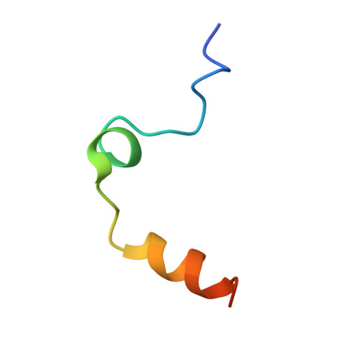
Thrombin light chain
A, C
33Homo sapiensMutation(s): 0 
Gene Names: F2
EC: 3.4.21.5
UniProt & NIH Common Fund Data Resources
Find proteins for P00734 (Homo sapiens)
Explore P00734 
Go to UniProtKB:  P00734
PHAROS:  P00734
GTEx:  ENSG00000180210 
Entity Groups  
Sequence Clusters30% Identity50% Identity70% Identity90% Identity95% Identity100% Identity
UniProt GroupP00734
Sequence Annotations
Expand
  • Reference Sequence
Find similar proteins by:  (by identity cutoff)  |  3D Structure
Entity ID: 2
MoleculeChains Sequence LengthOrganismDetailsImage
Thrombin heavy chain
B, D
259Homo sapiensMutation(s): 1 
Gene Names: F2
EC: 3.4.21.5
UniProt & NIH Common Fund Data Resources
Find proteins for P00734 (Homo sapiens)
Explore P00734 
Go to UniProtKB:  P00734
PHAROS:  P00734
GTEx:  ENSG00000180210 
Entity Groups  
Sequence Clusters30% Identity50% Identity70% Identity90% Identity95% Identity100% Identity
UniProt GroupP00734
Glycosylation
Glycosylation Sites: 1Go to GlyGen: P00734-1
Sequence Annotations
Expand
  • Reference Sequence
Oligosaccharides

Help

Entity ID: 3
MoleculeChains Length2D Diagram Glycosylation3D Interactions
2-acetamido-2-deoxy-beta-D-glucopyranose-(1-6)-2-acetamido-2-deoxy-beta-D-glucopyranose
E, F
2N-Glycosylation
Glycosylation Resources
GlyTouCan:  G35932AR
GlyCosmos:  G35932AR
GlyGen:  G35932AR
Experimental Data & Validation

Experimental Data

  • Method: X-RAY DIFFRACTION
  • Resolution: 1.70 Å
  • R-Value Free: 0.206 
  • R-Value Work: 0.181 
  • R-Value Observed: 0.182 
  • Space Group: P 21 21 21
Unit Cell:
Length ( Å )Angle ( ˚ )
a = 55.429α = 90
b = 81.333β = 90
c = 146.366γ = 90
Software Package:
Software NamePurpose
REFMACrefinement
HKL-2000data reduction
HKL-2000data scaling
MOLREPphasing

Structure Validation

View Full Validation Report


Entry History & Funding Information

Deposition Data


Funding OrganizationLocationGrant Number
National Institutes of Health/National Heart, Lung, and Blood Institute (NIH/NHLBI)United StatesHL049413
National Institutes of Health/National Heart, Lung, and Blood Institute (NIH/NHLBI)United StatesHL073813
National Institutes of Health/National Heart, Lung, and Blood Institute (NIH/NHLBI)United StatesHL112303

Revision History  (Full details and data files)

  • Version 1.0: 2016-07-13
    Type: Initial release
  • Version 1.1: 2016-08-10
    Changes: Database references
  • Version 1.2: 2017-09-27
    Changes: Author supporting evidence, Database references, Derived calculations
  • Version 1.3: 2019-12-04
    Changes: Author supporting evidence
  • Version 2.0: 2020-07-29
    Type: Remediation
    Reason: Carbohydrate remediation
    Changes: Atomic model, Data collection, Derived calculations, Structure summary
  • Version 2.1: 2023-09-27
    Changes: Data collection, Database references, Derived calculations, Refinement description, Structure summary
  • Version 2.2: 2024-10-30
    Changes: Structure summary