


Structural basis for substrate recognition and catalysis of a novel esterase E22 with a homoserine transacetylase-like fold
Zhang, Y., Yao, Q., Wang, P.To be published.
Experimental Data Snapshot
Starting Model: experimental
View more details
wwPDB Validation 3D Report Full Report

Entity ID: 1 | |||||
|---|---|---|---|---|---|
| Molecule | Chains | Sequence Length | Organism | Details | Image |
| Esterase E22 | 370 | uncultured bacterium | Mutation(s): 0 | 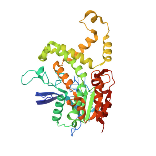 | |
| Length ( Å ) | Angle ( ˚ ) |
|---|---|
| a = 58.619 | α = 90 |
| b = 68.898 | β = 91.11 |
| c = 82.524 | γ = 90 |
| Software Name | Purpose |
|---|---|
| PHENIX | refinement |
| HKL-2000 | data reduction |
| HKL-2000 | data scaling |
| PHASER | phasing |


