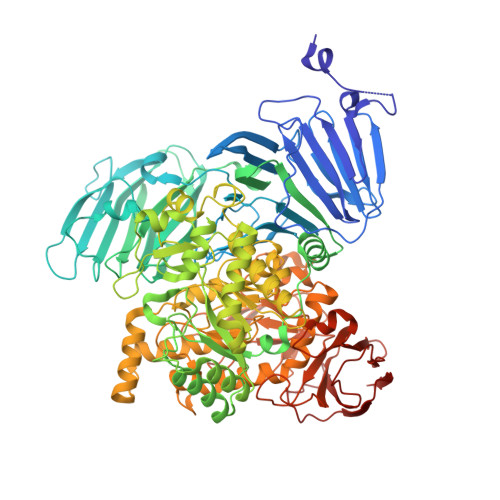
5JOV

Bacteroides ovatus Xyloglucan PUL GH31 with bound 5FIdoF


Experimental Data Snapshot

  • Method: X-RAY DIFFRACTION
  • Resolution: 1.50 Å
  • R-Value Free: 0.147 
  • R-Value Work: 0.107 
  • R-Value Observed: 0.109 

Starting Model: experimental
View more details

wwPDB Validation   3D Report Full Report


Ligand Structure Quality Assessment 


This is version 2.1 of the entry. See complete history


Literature

Structural dissection of a complex Bacteroides ovatus gene locus conferring xyloglucan metabolism in the human gut.

Hemsworth, G.R.Thompson, A.J.Stepper, J.Sobala, F.Coyle, T.Larsbrink, J.Spadiut, O.Goddard-Borger, E.D.Stubbs, K.A.Brumer, H.Davies, G.J.

(2016) Open Biol 6

  • DOI: https://doi.org/10.1098/rsob.160142
  • Primary Citation of Related Structures:  
    5JOU, 5JOV, 5JOW, 5JOX, 5JOY, 5JOZ, 5JP0

  • PubMed Abstract: 

    The human gastrointestinal tract harbours myriad bacterial species, collectively termed the microbiota, that strongly influence human health. Symbiotic members of our microbiota play a pivotal role in the digestion of complex carbohydrates that are otherwise recalcitrant to assimilation. Indeed, the intrinsic human polysaccharide-degrading enzyme repertoire is limited to various starch-based substrates; more complex polysaccharides demand microbial degradation. Select Bacteroidetes are responsible for the degradation of the ubiquitous vegetable xyloglucans (XyGs), through the concerted action of cohorts of enzymes and glycan-binding proteins encoded by specific xyloglucan utilization loci (XyGULs). Extending recent (meta)genomic, transcriptomic and biochemical analyses, significant questions remain regarding the structural biology of the molecular machinery required for XyG saccharification. Here, we reveal the three-dimensional structures of an α-xylosidase, a β-glucosidase, and two α-l-arabinofuranosidases from the Bacteroides ovatus XyGUL. Aided by bespoke ligand synthesis, our analyses highlight key adaptations in these enzymes that confer individual specificity for xyloglucan side chains and dictate concerted, stepwise disassembly of xyloglucan oligosaccharides. In harness with our recent structural characterization of the vanguard endo-xyloglucanse and cell-surface glycan-binding proteins, the present analysis provides a near-complete structural view of xyloglucan recognition and catalysis by XyGUL proteins.


  • Organizational Affiliation

    Department of Chemistry, York Structural Biology Laboratory, The University of York, Heslington, York YO10 5DD, UK.


Macromolecules
Find similar proteins by:  (by identity cutoff)  |  3D Structure
Entity ID: 1
MoleculeChains Sequence LengthOrganismDetailsImage
Alpha-xylosidase BoGH31A955Bacteroides ovatusMutation(s): 0 
Gene Names: BACOVA_02646
EC: 3.2.1.177
UniProt
Find proteins for A7LXT0 (Bacteroides ovatus (strain ATCC 8483 / DSM 1896 / JCM 5824 / BCRC 10623 / CCUG 4943 / NCTC 11153))
Explore A7LXT0 
Go to UniProtKB:  A7LXT0
Entity Groups  
Sequence Clusters30% Identity50% Identity70% Identity90% Identity95% Identity100% Identity
UniProt GroupA7LXT0
Sequence Annotations
Expand
  • Reference Sequence
Small Molecules
Ligands 4 Unique
IDChains Name / Formula / InChI Key2D Diagram3D Interactions
EPE
Query on EPE

Download Ideal Coordinates CCD File 
H [auth A]4-(2-HYDROXYETHYL)-1-PIPERAZINE ETHANESULFONIC ACID
C8 H18 N2 O4 S
JKMHFZQWWAIEOD-UHFFFAOYSA-N
B9D
Query on B9D

Download Ideal Coordinates CCD File 
I [auth A]5-fluoro-alpha-L-idopyranose
C6 H11 F O6
YQZCKDSOGGIGPL-DVKNGEFBSA-N
EDO
Query on EDO

Download Ideal Coordinates CCD File 
C [auth A],
D [auth A],
E [auth A],
F [auth A],
G [auth A]
1,2-ETHANEDIOL
C2 H6 O2
LYCAIKOWRPUZTN-UHFFFAOYSA-N
NI
Query on NI

Download Ideal Coordinates CCD File 
B [auth A]NICKEL (II) ION
Ni
VEQPNABPJHWNSG-UHFFFAOYSA-N
Experimental Data & Validation

Experimental Data

  • Method: X-RAY DIFFRACTION
  • Resolution: 1.50 Å
  • R-Value Free: 0.147 
  • R-Value Work: 0.107 
  • R-Value Observed: 0.109 
  • Space Group: P 21 21 21
Unit Cell:
Length ( Å )Angle ( ˚ )
a = 76.906α = 90
b = 108.448β = 90
c = 144.853γ = 90
Software Package:
Software NamePurpose
REFMACrefinement
XDSdata reduction
Aimlessdata scaling
MOLREPphasing

Structure Validation

View Full Validation Report


Ligand Structure Quality Assessment 


Entry History & Funding Information

Deposition Data


Funding OrganizationLocationGrant Number
Biotechnology and Biological Sciences Research CouncilUnited KingdomBB/K00591X/1
Biotechnology and Biological Sciences Research CouncilUnited KingdomBB/I014802/1
European Research CouncilUnited Kingdom322942

Revision History  (Full details and data files)

  • Version 1.0: 2016-08-10
    Type: Initial release
  • Version 1.1: 2017-08-30
    Changes: Author supporting evidence
  • Version 2.0: 2020-07-29
    Type: Remediation
    Reason: Carbohydrate remediation
    Changes: Atomic model, Derived calculations, Structure summary
  • Version 2.1: 2024-01-10
    Changes: Data collection, Database references, Refinement description, Structure summary