5JXF | pdb_00005jxf

Crystal structure of Flavobacterium psychrophilum DPP11 in complex with dipeptide Arg-Asp


Experimental Data Snapshot

  • Method: X-RAY DIFFRACTION
  • Resolution: 2.10 Å
  • R-Value Free: 
    0.244 (Depositor), 0.245 (DCC) 
  • R-Value Work: 
    0.196 (Depositor), 0.201 (DCC) 
  • R-Value Observed: 
    0.198 (Depositor) 

Starting Model: experimental
View more details

wwPDB Validation 3D Report Full Report

Validation slider image for 5JXF

Ligand Structure Quality Assessment 


This is version 1.2 of the entry. See complete history

Literature

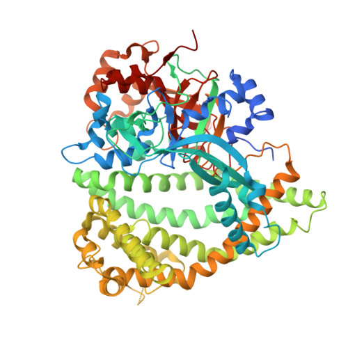
Bacterial protease uses distinct thermodynamic signatures for substrate recognition.

Bezerra, G.A.Ohara-Nemoto, Y.Cornaciu, I.Fedosyuk, S.Hoffmann, G.Round, A.Marquez, J.A.Nemoto, T.K.Djinovic-Carugo, K.

(2017) Sci Rep 7: 2848-2848

  • DOI: https://doi.org/10.1038/s41598-017-03220-y
  • Primary Citation Related Structures: 
    5JWF, 5JWG, 5JWI, 5JXF, 5JXK, 5JXP, 5JY0

  • PubMed Abstract: 

    Porphyromonas gingivalis and Porphyromonas endodontalis are important bacteria related to periodontitis, the most common chronic inflammatory disease in humans worldwide. Its comorbidity with systemic diseases, such as type 2 diabetes, oral cancers and cardiovascular diseases, continues to generate considerable interest. Surprisingly, these two microorganisms do not ferment carbohydrates; rather they use proteinaceous substrates as carbon and energy sources. However, the underlying biochemical mechanisms of their energy metabolism remain unknown. Here, we show that dipeptidyl peptidase 11 (DPP11), a central metabolic enzyme in these bacteria, undergoes a conformational change upon peptide binding to distinguish substrates from end products. It binds substrates through an entropy-driven process and end products in an enthalpy-driven fashion. We show that increase in protein conformational entropy is the main-driving force for substrate binding via the unfolding of specific regions of the enzyme ("entropy reservoirs"). The relationship between our structural and thermodynamics data yields a distinct model for protein-protein interactions where protein conformational entropy modulates the binding free-energy. Further, our findings provide a framework for the structure-based design of specific DPP11 inhibitors.


  • Organizational Affiliation
    • Department of Structural & Computational Biology, Max F. Perutz Laboratories, University of Vienna, Vienna Biocenter, Vienna Biocenter Campus 5, A-1030, Vienna, Austria. gustavo.bezerra@univie.ac.at.

Macromolecule Content 

  • Total Structure Weight: 331.46 kDa 
  • Atom Count: 22,036 
  • Modeled Residue Count: 2,753 
  • Deposited Residue Count: 2,924 
  • Unique protein chains: 1

Macromolecules

Find similar proteins by:|  3D Structure
Entity ID: 1
MoleculeChains  Sequence LengthOrganismDetailsImage
Asp/Glu-specific dipeptidyl-peptidase
A, B, C, D
731Flavobacterium psychrophilumMutation(s): 0 
Gene Names: dpp11FP0382
EC: 3.4.14
UniProt
Find proteins for A6GWM2 (Flavobacterium psychrophilum (strain ATCC 49511 / DSM 21280 / CIP 103535 / JIP02/86))
Explore A6GWM2 
Go to UniProtKB:  A6GWM2
Entity Groups
Sequence Clusters30% Identity50% Identity70% Identity90% Identity95% Identity100% Identity
UniProt GroupA6GWM2
Sequence Annotations
Expand
Reference Sequence

Small Molecules

Ligands 4 Unique
IDChains Name / Formula / InChI Key2D Diagram3D Interactions
ARG

Query on ARG


Download:Ideal Coordinates CCD File
E [auth A],
G [auth B]
ARGININE
C6 H15 N4 O2
ODKSFYDXXFIFQN-BYPYZUCNSA-O
ASP

Query on ASP


Download:Ideal Coordinates CCD File
F [auth A],
H [auth B]
ASPARTIC ACID
C4 H7 N O4
CKLJMWTZIZZHCS-REOHCLBHSA-N
CL

Query on CL


Download:Ideal Coordinates CCD File
I [auth C],
J [auth D],
K [auth D]
CHLORIDE ION
Cl
VEXZGXHMUGYJMC-UHFFFAOYSA-M
NA

Query on NA


Download:Ideal Coordinates CCD File
L [auth D]SODIUM ION
Na
FKNQFGJONOIPTF-UHFFFAOYSA-N

Experimental Data & Validation

Experimental Data

  • Method: X-RAY DIFFRACTION
  • Resolution: 2.10 Å
  • R-Value Free:  0.244 (Depositor), 0.245 (DCC) 
  • R-Value Work:  0.196 (Depositor), 0.201 (DCC) 
  • R-Value Observed: 0.198 (Depositor) 
Space Group: P 1 21 1
Unit Cell:
Length ( Å )Angle ( ˚ )
a = 126.051α = 90
b = 70.689β = 97.26
c = 191.598γ = 90
Software Package:
Software NamePurpose
PHENIXrefinement
SCALAdata scaling
PDB_EXTRACTdata extraction
XDSdata reduction
PHASERphasing

Structure Validation

View Full Validation Report


Ligand Structure Quality Assessment 


Entry History 

Deposition Data

Revision History  (Full details and data files)

  • Version 1.0: 2017-06-14
    Type: Initial release
  • Version 1.1: 2017-06-21
    Changes: Database references
  • Version 1.2: 2024-01-10
    Changes: Data collection, Database references, Refinement description