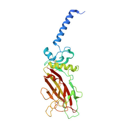
5K31

Crystal structure of Human fibrillar procollagen type I C-propeptide Homo-trimer


Experimental Data Snapshot

  • Method: X-RAY DIFFRACTION
  • Resolution: 2.20 Å
  • R-Value Free: 0.238 
  • R-Value Work: 0.196 
  • R-Value Observed: 0.198 

Starting Model: experimental
View more details

wwPDB Validation   3D Report Full Report


This is version 1.2 of the entry. See complete history


Literature

Structural basis of homo- and heterotrimerization of collagen I.

Sharma, U.Carrique, L.Vadon-Le Goff, S.Mariano, N.Georges, R.N.Delolme, F.Koivunen, P.Myllyharju, J.Moali, C.Aghajari, N.Hulmes, D.J.

(2017) Nat Commun 8: 14671-14671

  • DOI: https://doi.org/10.1038/ncomms14671
  • Primary Citation of Related Structures:  
    5K31

  • PubMed Abstract: 

    Fibrillar collagen molecules are synthesized as precursors, procollagens, with large propeptide extensions. While a homotrimeric form (three α1 chains) has been reported in embryonic tissues as well as in diseases (cancer, fibrosis, genetic disorders), collagen type I usually occurs as a heterotrimer (two α1 chains and one α2 chain). Inside the cell, the role of the C-terminal propeptides is to gather together the correct combination of three α chains during molecular assembly, but how this occurs for different forms of the same collagen type is so far unknown. Here, by structural and mutagenic analysis, we identify key amino acid residues in the α1 and α2 C-propeptides that determine homo- and heterotrimerization. A naturally occurring mutation in one of these alters the homo/heterotrimer balance. These results show how the C-propeptide of the α2 chain has specifically evolved to permit the appearance of heterotrimeric collagen I, the major extracellular building block among the metazoa.


  • Organizational Affiliation

    Molecular Microbiology and Structural Biochemistry Unit, UMR 5086 CNRS - University of Lyon 1, 7 passage du Vercors, F-69367 Lyon, France.


Macromolecules
Find similar proteins by:  (by identity cutoff)  |  3D Structure
Entity ID: 1
MoleculeChains Sequence LengthOrganismDetailsImage
Collagen alpha-1(I) chain
A, B, C, D, E
A, B, C, D, E, F
255Homo sapiensMutation(s): 2 
Gene Names: COL1A1
UniProt & NIH Common Fund Data Resources
Find proteins for P02452 (Homo sapiens)
Explore P02452 
Go to UniProtKB:  P02452
PHAROS:  P02452
GTEx:  ENSG00000108821 
Entity Groups  
Sequence Clusters30% Identity50% Identity70% Identity90% Identity95% Identity100% Identity
UniProt GroupP02452
Sequence Annotations
Expand
  • Reference Sequence
Small Molecules
Ligands 3 Unique
IDChains Name / Formula / InChI Key2D Diagram3D Interactions
GOL
Query on GOL

Download Ideal Coordinates CCD File 
H [auth A],
K [auth B],
L [auth B],
M [auth B],
S [auth E]
GLYCEROL
C3 H8 O3
PEDCQBHIVMGVHV-UHFFFAOYSA-N
CA
Query on CA

Download Ideal Coordinates CCD File 
G [auth A]
I [auth B]
O [auth C]
P [auth D]
R [auth E]
G [auth A],
I [auth B],
O [auth C],
P [auth D],
R [auth E],
T [auth F]
CALCIUM ION
Ca
BHPQYMZQTOCNFJ-UHFFFAOYSA-N
CL
Query on CL

Download Ideal Coordinates CCD File 
J [auth B],
N [auth B],
Q [auth D]
CHLORIDE ION
Cl
VEXZGXHMUGYJMC-UHFFFAOYSA-M
Experimental Data & Validation

Experimental Data

  • Method: X-RAY DIFFRACTION
  • Resolution: 2.20 Å
  • R-Value Free: 0.238 
  • R-Value Work: 0.196 
  • R-Value Observed: 0.198 
  • Space Group: P 1 21 1
Unit Cell:
Length ( Å )Angle ( ˚ )
a = 74.82α = 90
b = 149.63β = 101.68
c = 105.95γ = 90
Software Package:
Software NamePurpose
REFMACrefinement
XDSdata reduction
XSCALEdata scaling
PHASERphasing

Structure Validation

View Full Validation Report


Entry History & Funding Information

Deposition Data


Funding OrganizationLocationGrant Number
French National Research AgencyFrance2010-BLAN-1526-01

Revision History  (Full details and data files)

  • Version 1.0: 2017-03-22
    Type: Initial release
  • Version 1.1: 2017-08-30
    Changes: Author supporting evidence
  • Version 1.2: 2024-01-10
    Changes: Data collection, Database references, Derived calculations, Refinement description