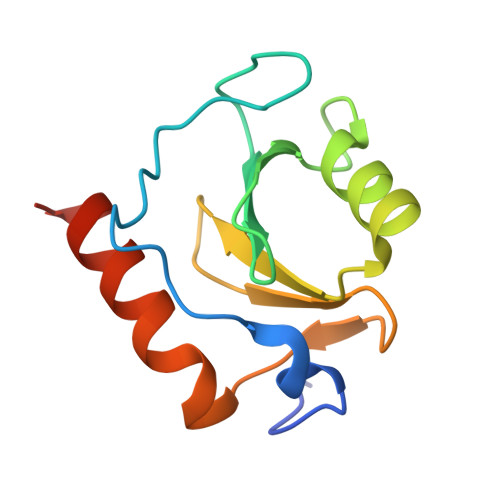
5KH7

Crystal structure of fragment (3-[6-Oxo-3-(3-pyridinyl)-1(6H)-pyridazinyl]propanoic acid) bound in the ubiquitin binding pocket of the HDAC6 zinc-finger domain


Experimental Data Snapshot

  • Method: X-RAY DIFFRACTION
  • Resolution: 1.70 Å
  • R-Value Free: 0.183 
  • R-Value Work: 0.146 
  • R-Value Observed: 0.148 

Starting Model: experimental
View more details

wwPDB Validation   3D Report Full Report


Ligand Structure Quality Assessment 


This is version 1.3 of the entry. See complete history


Literature

Small Molecule Antagonists of the Interaction between the Histone Deacetylase 6 Zinc-Finger Domain and Ubiquitin.

Harding, R.J.Ferreira de Freitas, R.Collins, P.Franzoni, I.Ravichandran, M.Ouyang, H.Juarez-Ornelas, K.A.Lautens, M.Schapira, M.von Delft, F.Santhakumar, V.Arrowsmith, C.H.

(2017) J Med Chem 60: 9090-9096

  • DOI: https://doi.org/10.1021/acs.jmedchem.7b00933
  • Primary Citation of Related Structures:  
    5B8D, 5KH3, 5KH7, 5KH9, 5WPB

  • PubMed Abstract: 

    Inhibitors of HDAC6 have attractive potential in numerous cancers. HDAC6 inhibitors to date target the catalytic domains, but targeting the unique zinc-finger ubiquitin-binding domain (Zf-UBD) of HDAC6 may be an attractive alternative strategy. We developed X-ray crystallography and biophysical assays to identify and characterize small molecules capable of binding to the Zf-UBD and competing with ubiquitin binding. Our results revealed two adjacent ligand-able pockets of HDAC6 Zf-UBD and the first functional ligands for this domain.


  • Organizational Affiliation

    Structural Genomics Consortium, University of Toronto , MaRS South Tower, Suite 700, 101 College Street, Toronto, Ontario M5G 1L7, Canada.


Macromolecules
Find similar proteins by:  (by identity cutoff)  |  3D Structure
Entity ID: 1
MoleculeChains Sequence LengthOrganismDetailsImage
Histone deacetylase 6107Homo sapiensMutation(s): 0 
Gene Names: HDAC6KIAA0901JM21
EC: 3.5.1.98 (PDB Primary Data), 3.5.1 (UniProt)
UniProt & NIH Common Fund Data Resources
Find proteins for Q9UBN7 (Homo sapiens)
Explore Q9UBN7 
Go to UniProtKB:  Q9UBN7
PHAROS:  Q9UBN7
GTEx:  ENSG00000094631 
Entity Groups  
Sequence Clusters30% Identity50% Identity70% Identity90% Identity95% Identity100% Identity
UniProt GroupQ9UBN7
Sequence Annotations
Expand
  • Reference Sequence
Small Molecules
Ligands 3 Unique
IDChains Name / Formula / InChI Key2D Diagram3D Interactions
6T7
Query on 6T7

Download Ideal Coordinates CCD File 
E [auth A]3-(6-oxidanylidene-3-pyridin-3-yl-pyridazin-1-yl)propanoic acid
C12 H11 N3 O3
FPHNPEYKDLCEEE-UHFFFAOYSA-N
ZN
Query on ZN

Download Ideal Coordinates CCD File 
B [auth A],
C [auth A],
D [auth A]
ZINC ION
Zn
PTFCDOFLOPIGGS-UHFFFAOYSA-N
UNX
Query on UNX

Download Ideal Coordinates CCD File 
F [auth A]
G [auth A]
H [auth A]
I [auth A]
J [auth A]
F [auth A],
G [auth A],
H [auth A],
I [auth A],
J [auth A],
K [auth A],
L [auth A],
M [auth A],
N [auth A],
O [auth A],
P [auth A],
Q [auth A],
R [auth A],
S [auth A],
T [auth A]
UNKNOWN ATOM OR ION
X
Experimental Data & Validation

Experimental Data

Unit Cell:
Length ( Å )Angle ( ˚ )
a = 40.719α = 90
b = 43.776β = 90
c = 55.924γ = 90
Software Package:
Software NamePurpose
REFMACrefinement
Aimlessdata scaling
PDB_EXTRACTdata extraction
XDSdata reduction

Structure Validation

View Full Validation Report


Ligand Structure Quality Assessment 


Entry History 

Revision History  (Full details and data files)

  • Version 1.0: 2016-07-27
    Type: Initial release
  • Version 1.1: 2018-03-21
    Changes: Database references, Derived calculations
  • Version 1.2: 2019-02-27
    Changes: Data collection, Database references
  • Version 1.3: 2023-09-27
    Changes: Data collection, Database references, Refinement description