Discovery of imidazopyridazines as potent Pim-1/2 kinase inhibitors.
Wurz, R.P., Sastri, C., D'Amico, D.C., Herberich, B., Jackson, C.L., Pettus, L.H., Tasker, A.S., Wu, B., Guerrero, N., Lipford, J.R., Winston, J.T., Yang, Y., Wang, P., Nguyen, Y., Andrews, K.L., Huang, X., Lee, M.R., Mohr, C., Zhang, J.D., Reid, D.L., Xu, Y., Zhou, Y., Wang, H.L.(2016) Bioorg Med Chem Lett 26: 5580-5590
- PubMed: 27769621 Search on PubMed
- DOI: https://doi.org/10.1016/j.bmcl.2016.09.067
- Primary Citation Related Structures:
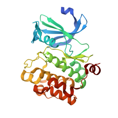
5KZI - PubMed Abstract:
High levels of Pim expression have been implicated in several hematopoietic and solid tumor cancers, suggesting that inhibition of Pim signaling could provide patients with therapeutic benefit. Herein, we describe our progress towards this goal using a screening hit (rac-1) as a starting point. Modification of the indazole ring resulted in the discovery of a series of imidazopyridazine-based Pim inhibitors exemplified by compound 22m, which was found to be a subnanomolar inhibitor of the Pim-1 and Pim-2 isoforms (IC 50 values of 0.024nM and 0.095nM, respectively) and to potently inhibit the phosphorylation of BAD in a cell line that expresses high levels of all Pim isoforms, KMS-12-BM (IC 50 =28nM). Profiling of Pim-1 and Pim-2 expression levels in a panel of multiple myeloma cell lines and correlation of these data with the potency of compound 22m in a proliferation assay suggests that Pim-2 inhibition would be advantageous for this indication.
- Department of Therapeutic Discovery, Amgen Inc., One Amgen Center Drive, Thousand Oaks, CA 91320-1799, USA. Electronic address: rwurz@amgen.com.
Organizational Affiliation: